


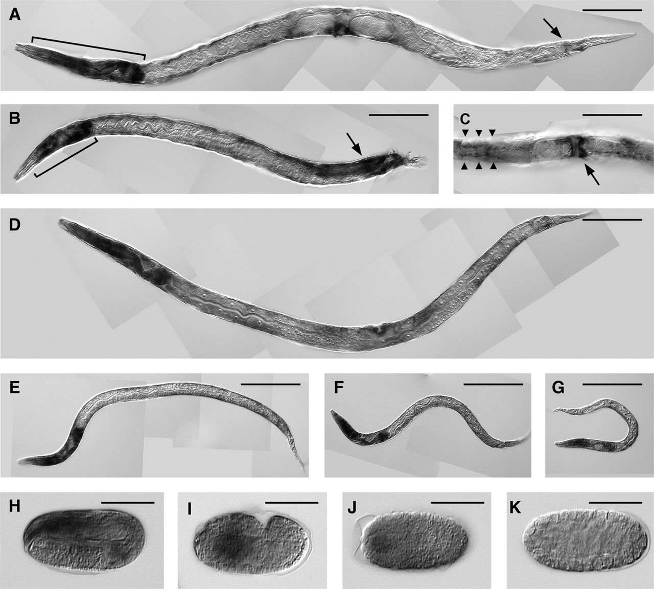
FIGURE 1.
WISH analysis for detection of mir-1 miRNA. Images show an adult hermaphrodite (A), an adult male (B), and a ventral view of the vulva of an adult hermaphrodite (C). The brackets designate the pharynx (A,B). The arrows indicate the anal depressor muscle (A), the diagonal muscle (B), and the vulval muscle (C). The arrowheads show the body-wall muscle (C). Larvae are at the L4 (D), L3 (E), L2 (F), and L1 (G) stages. Embryos are at the fourfold (H), 1.5-fold (I), 500-cell (J), and 200-cell (K) stages. Scale bars, 100 µm (A–G) and 30 µm (H–K).




