


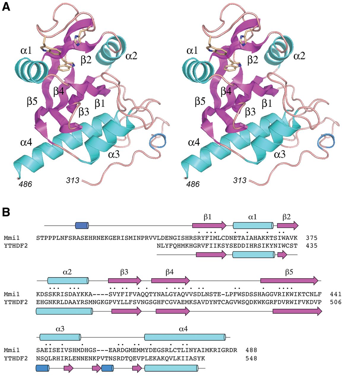
Structure of the Mmi1 YTH domain. (A) Stereo view of the tertiary structure of Mmi1(311–488), depicted as a ribbon model composed of five β strands (magenta), four α helices (cyan), and one 310 helix (blue). N- and C-terminal amino acids are numbered in italics. The three amino acids corresponding to the aromatic cage that binds to m6A in other YTH proteins are shown as stick models. (B) The Mmi1 and YTHDF2 primary structures are aligned according to DALI. Gaps in the alignment are indicated by dashes. Positions of amino acid side chain identity/similarity are indicated by (•) above the sequence. The secondary structure elements of Mmi1 and YTHDF2 (colored as in panel A) are shown above and below the respective amino acid sequences.




