


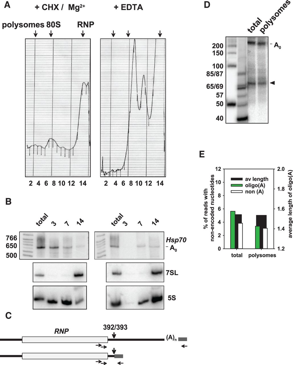
Oligoadenylated 3′ decay intermediates are present on polysomes. (A) Cytoplasmic extract was prepared from S2 cells expressing the Hsp70 reporter and fractioned by sucrose gradient centrifugation (see Materials and Methods). The panel on the left shows the UV trace of a sample from cycloheximide-treated cells analyzed in the presence of Mg2+; the panel on the right shows the UV trace of a comparable sample from cells not treated with cycloheximide and mixed with EDTA to disrupt ribosomes. (B) RNA was isolated from fractions 3, 7, and 14 of the gradients shown in A and analyzed by Northern blotting. 7SL RNA and 5S RNA were used as markers for the RNP fraction and ribosomes, respectively. (C) An RT-PCR procedure as outlined in Figure 1 was used to generate sequencing libraries. A single nested pair of upstream primers was used. The scheme shows how the two main products were generated by adaptors ligated to the completely deadenylated full-length RNA and to the 3′ decay intermediate at nt 392/393. (D) For analytical purposes, the same PCR was carried out with [α-32P]-dCTP, and the products were separated on a denaturing polyacrylamide gel and analyzed by phosphorimaging. Marker sizes are indicated on the left. The main product corresponds to the size expected for amplification of full-length RNA. The second most abundant product (arrowhead) corresponds to the size expected for amplification of the fragment terminating at nt 392/393. (E) Total RNA and polysomal RNA was analyzed by Illumina sequencing. Read numbers obtained were between 303,000 and 518,000 per sequencing library. The data were averaged from two independent experiments.




