


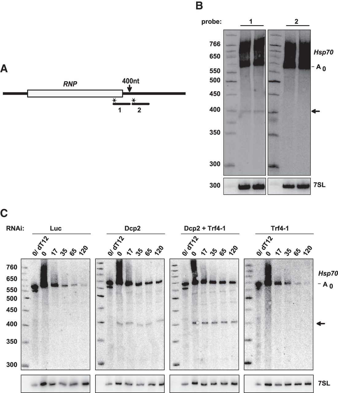
An intermediate of 3′ decay accumulates upon knockdown of Trf4-1. (A) The scheme of the reporter RNA and probes used for the Northern blot. The arrow indicates the position of the approximate 3′ end of the main 3′ decay intermediate visible in Northern blots. (B) S2 cells expressing the Hsp70 reporter were treated with Dcp2 dsRNA. Total RNA was isolated and analyzed by Northern blotting with the probes depicted in A. The ∼400 nt fragment (arrow) was detectable only with probe 1, but not with probe 2. Identification of the deadenylated RNA species is based on comparison to RNaseH/oligo(dT) digestions such as shown in C. (C) S2 cells expressing the Hsp70 reporter were treated with dsRNA against Dcp2 and/or Trf4-1 as indicated. Reporter transcription was induced and inhibited by actinomycin D after 60 min. Total RNA was isolated at the indicated time points after actinomycin D addition and analyzed by Northern blotting with a reporter-specific probe. For quantitation, the intensity of the ∼400 nt 3′ decay intermediate (arrow) was normalized to the intensity of the fully deadenylated decay intermediate (A0).




