


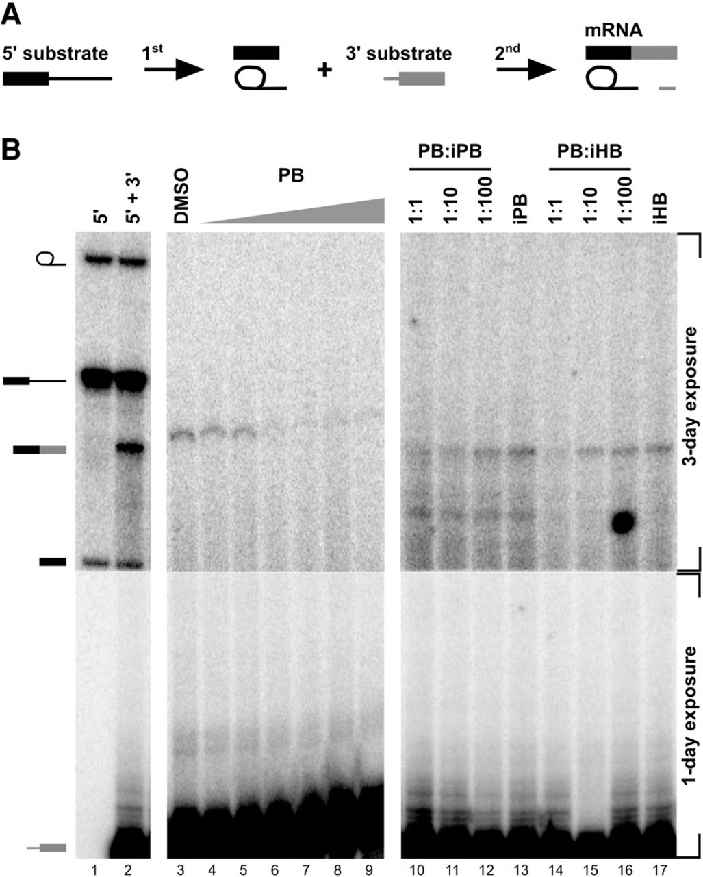
Bimolecular assay shows that PB inhibits exon ligation. (A) Schematic of the bimolecular exon ligation reaction. The 5′ substrate consists of a 5′ exon and intron containing the branch point and PYT and is capable of 1st step splicing chemistry to produce splicing intermediates. The 3′ substrate consists of a 3′ splice site and 3′ exon and is capable of 2nd step chemistry to produce mRNA when added in trans. To test for inhibition, PB is added with the 3′ substrate. (B) Denaturing gel analysis of in vitro bimolecular exon ligation reactions. Identities of bands are schematized to the left as (from top to bottom) lariat intermediate, 5′ substrate, mRNA, 5′ exon, and 3′ exon. A 3-d exposure is shown for the top region of the gel, while the bottom region is a 1-d exposure. In lanes 1 and 2, both 5′ and 3′ substrates are radiolabeled. In lanes 3–17, only the 3′ substrate is radiolabeled. In lanes 4–9, increasing concentrations of PB (2) (0.1, 1, 2, 5, 10, 100 µM) were included with the 3′ substrate. In lanes 10–12 and 14–16, active compound was added at 2 µM and increasing concentrations of inactive compounds were added at the indicated ratio. In lanes 13 and 17, inactive compound is at 200 µM.




