


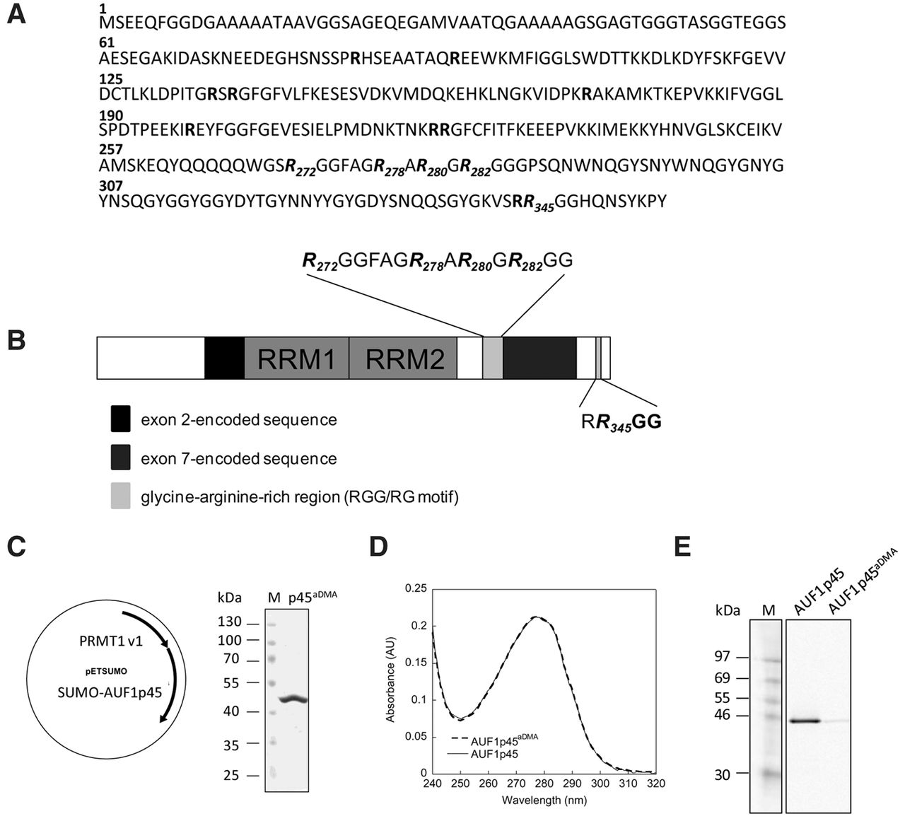
Identification of methylation sites; production and purification of methylated, recombinant AUF1 p45. (A) Amino acid sequence of AUF1 p45. All arginine residues in the sequence are highlighted in bold. The dimethylated arginines at positions 272, 278, 280, 282, and 345 that were identified by MALDI-TOF-MS after digestion with trypsin and chymotrypsin are highlighted in italics. (B) Schematic drawing of the domain organization of AUF1 p45. The RNA recognition motifs (RRM) are indicated as well as isoform-specific sequences encoded by exon 2 (black) and exon 7 (dark gray). The glycine-arginine-rich region (RGG/RG motif) within the C terminus of AUF1 p45 is indicated in light gray; the dimethylated arginines at positions 272, 278, 280, 282, and 345 that were identified by MALDI-TOF-MS are emphasized. (C) Schematic drawing of the bicistronic vector that was used to coexpress PRMT1 v1 and SUMO-AUF1 p45 in E. coli. The methylated AUF1 p45 (AUF1 p45aDMA) was purified as described in Materials and Methods. About 2 µg of purified AUF1 p45aDMA was analyzed on a Coomassie blue-stained SDS–PAGE in comparison to protein markers. (D) Absorption spectra of AUF1 p45 and AUF1 p45aDMA that were recombinantly expressed in and purified from E. coli. The ratio of the absorbance at 280 and 260 nm was about 1.8 for both protein preparations, indicating no contamination with nucleic acids. (E) In vitro methylation assay performed with 5 pmol purified PRMT1 v1 and 13 pmol of recombinant, nonmethylated AUF1 p45 and 13 pmol of recombinant, methylated AUF1 p45aDMA, respectively, using 400 pmol of [S-14C] adenosylmethionine as a substrate. The samples were taken after 2 h and analyzed by SDS–PAGE and the methylation state determined by phosphorimaging. One representative experiment is shown.




