


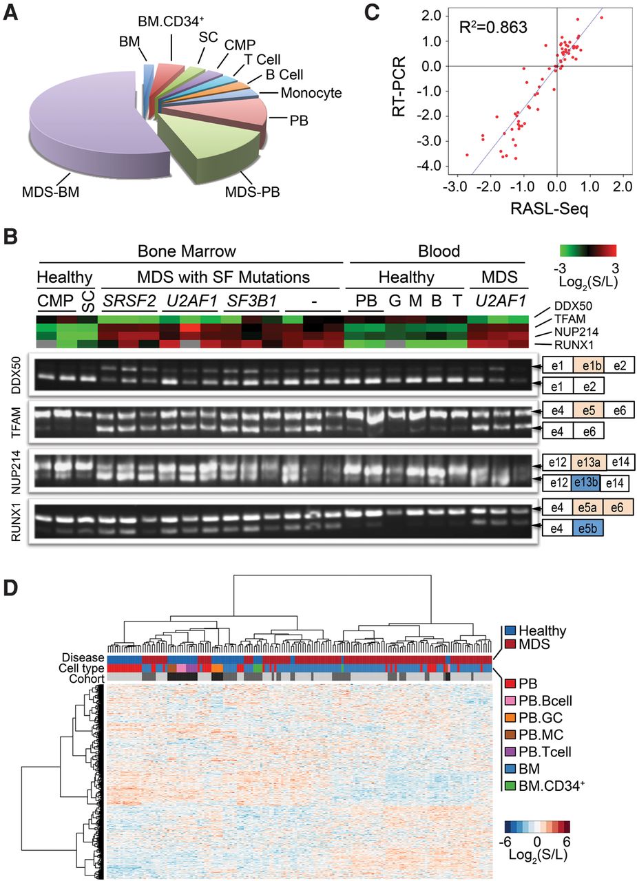
Characterization of lineage commitment and disease status by alternative splicing. RASL-seq was applied to 115 MDS samples and 54 samples from healthy volunteers to assess global pre-mRNA splicing. (A) Pie chart showing type and origin of investigated samples. Bone marrow (BM); peripheral blood (PB); common myeloid progenitor cells (CMP); stem cell (SC). (B) RT-PCR validation of four events across multiple samples with different sample origins, different splicing factor mutations from MDS patients, and healthy volunteers. (Top) Heatmap view of RASL-seq data. (Bottom) Corresponding RT-PCR products for validation. (C) Scatter plot of RASL-seq versus RT-PCR validated data. (S) Short isoform; (L) long isoform. (D) Global view of RASL-seq data [normalized Log2 (short isoform/long isoform)] using unsupervised hierarchical clustering. Monocyte (MC); granulocyte (GC); bone marrow (BM). Light gray, gray, and dark gray colored bars represent three separate sample cohorts.




