


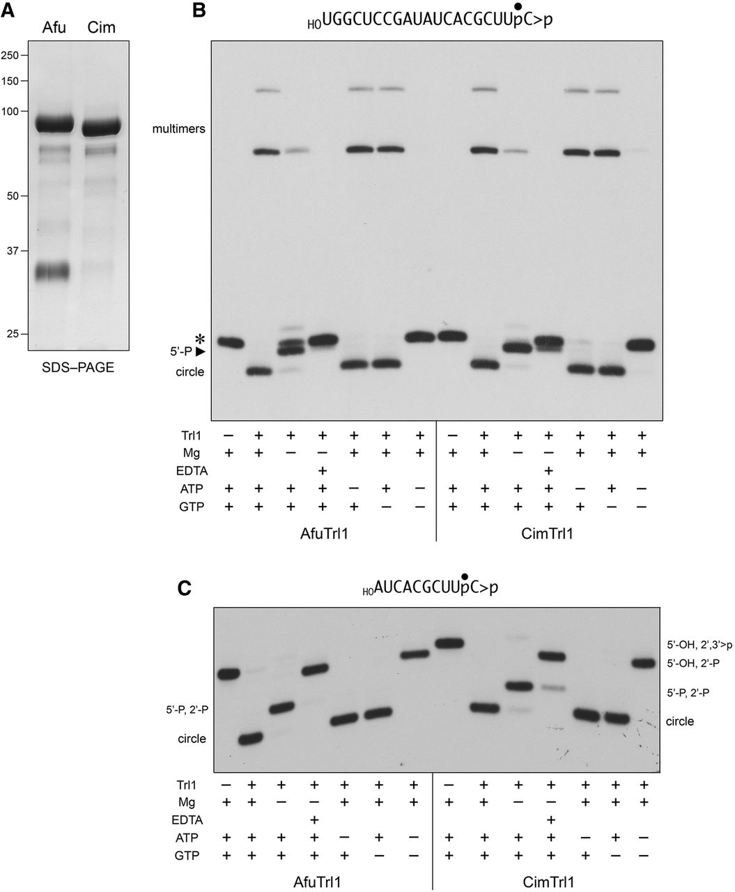
RNA end-joining activities of recombinant AfuTrl1 and CimTrl1. (A) Aliquots (5 µg) of the recombinant AfuTrl1 and CimTrl1 preparations were analyzed by SDS-PAGE. The Coomassie blue-stained gel is shown. The positions and sizes (kDa) of marker proteins are indicated on the left. (B,C) AfuTrl1 and CimTrl1 were reacted with a 3′ 32P-labeled 20-mer HORNA>p substrate (depicted at the top of panel B with the 32P-label denoted by •) or a 3′ 32P-labeled 10-mer HORNA>p substrate (depicted at the top of panel C). Complete reaction mixtures (20 µL) containing 50 mM Tris–HCl (pH 7.5), 2 mM DTT, 10 mM MgCl2, 100 µM ATP, 100 µM GTP, 20 nM HORNA>p, and 50 nM Trl1 were incubated for 5 min at 22°C. Individual reaction components were included (+) or omitted (−) as specified below the lanes; mixtures lacking 10 mM MgCl2 were supplemented with 10 mM EDTA where indicated by (+). The reactions were quenched with formamide/EDTA and the products were analyzed by urea–PAGE. Autoradiographs of the gels are shown. The positions and identities of the HORNA>p substrate (*), 5′-phosphorylated kinase product (5′-P), and ligation products (circle and multimers) are indicated on the left in panel B. The positions and identities of the input substrate and healed intermediates (specified by their termini), and circular ligation product are indicated on the left and right in panel C.




