


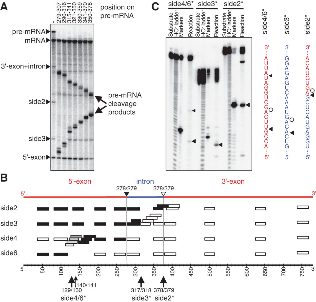
Identification of side products from the in vitro splicing reaction. (A) Phosphorimage of a denaturing PAGE separation of spliceozyme reaction products with internally 32P-labeled CAT pre-mRNA. After the reaction, the products were annealed with DNA oligonucleotides and treated with RNase H. The disappearance of a band showed that the DNA oligonucleotides had complementarity to the band's RNA. The positions covered by seven representative DNA oligonucleotides are shown above the gel image. These positions extend over the entire intron sequence. (B) Schematic of CAT pre-mRNA with the position of DNA oligonucleotides indicated. Effects of the DNA oligonucleotides on side product bands side2, side3, side4, and side6 are indicated by filled rectangles (band disappears) or empty rectangles (band is unaffected). Gray rectangles indicate partial disappearance. The position of 5′-exon (red), intron (blue), and 3′-exon (red) in the pre-mRNA are indicated on top. The nucleotide position is given on the bottom. The 5′-splice site is denoted by a black triangle; the 3′-splice site by an open triangle. Cleavage sites with single-nucleotide resolution (see (C)) are given on the bottom. (C) Identification of the cleavage sites at single-nucleotide resolution, using short substrate fragments with 5′-[32P] radiolabel. The sequences at which cleavage occurred are shown on the right, with triangles denoting cleavage sites by the spliceozyme and circles denoting the cleavage position by the DNAzyme to generate the marker. The resulting cleavage sites are annotated at the bottom of sub-figure (B).




