


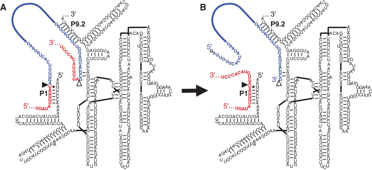
FIGURE 1.
Secondary structure of spliceozyme used as parent in the evolution. The spliceozyme (black) base pairs with the CAT pre-mRNA (red/blue) by forming duplexes P1 and P9.2. (A) Before the splicing reaction, the pre-mRNA contains the 5′-exon (red), a 100-nt intron (blue), and the 3′-exon (red). (B) After the splicing reaction, the 5′-exon and 3′-exon are joined to form CAT mRNA (red), and the intron (blue) is excised. The 5′-splice site is indicated by a filled triangle; the 3′-splice site is indicated by an open triangle. Note that the intron 5′ terminus is extended by an external guanosine during the reaction.




