


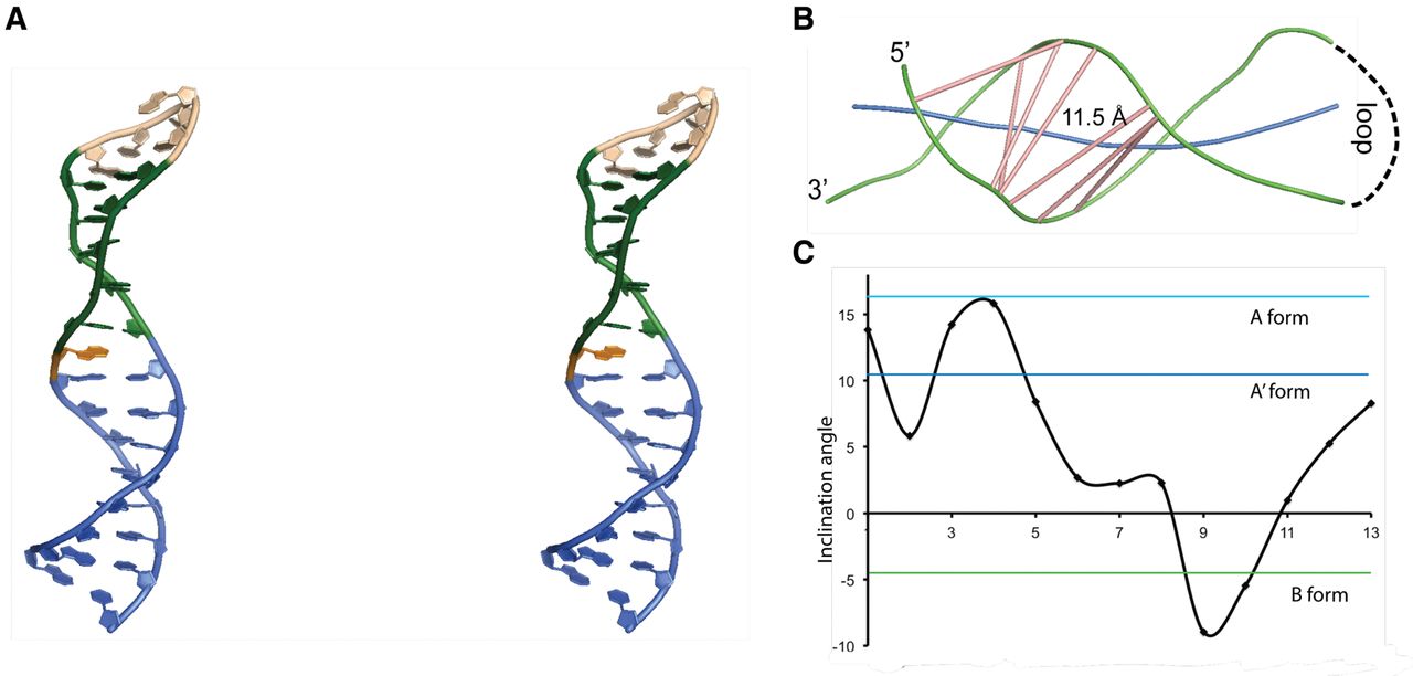
(A) Stereo view of a representative structure from the bundle of 10 lowest energy structures of the SOLE RNA (5a17.pdb). Blue, PS stem comprising nucleotides 1–8 and 25–32; green, base pairs in the MSL region, comprising nucleotides 9–13 and 19–23; pink, loop; orange, A24. (B) Representation of the major groove width of the SOLE RNA. Green, backbone splines; blue, RNA axis; pink, major groove vectors. The groove width is measured between the spline curves running through the phosphorus atoms. This width is reduced by 5.8 Å to account for the width of the phosphorus backbone. The figure was generated with the program CURVES+. (C) Base pair inclination angle with respect to the stem axis at each nucleotide position.




