


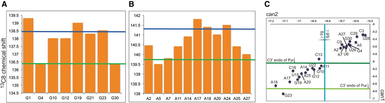
FIGURE 3.
(A,B) Chemical shifts of the 13C8 of G's (A) and A's (B). The green and blue lines indicate the average chemical shifts in helical and disordered regions, respectively, as defined in Fares et al. (2007). (C) Plot of the can1 and can2 coordinates for the riboses of the SOLE RNA (Ohlenschlager et al. 2008). Green lines indicate the minimum can1 for riboses of pyrimidines (dark green) and purines (light green) in the C3′ endo conformation. The blue line indicates the minimum can2 compatible with a g/g conformation of the γ dihedral angle.




