


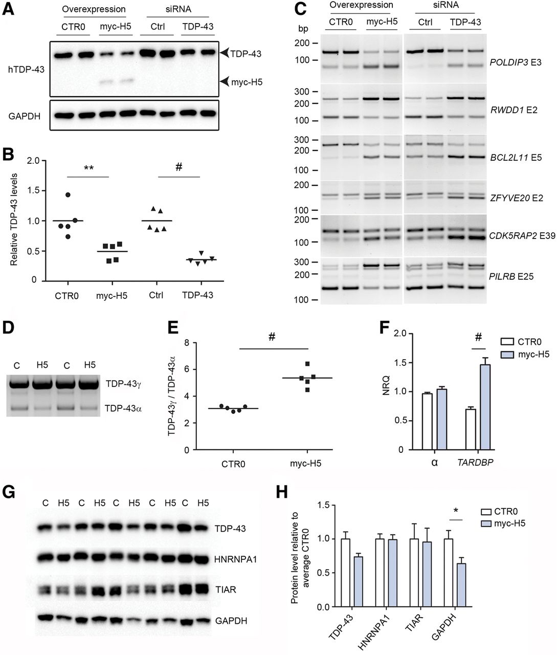
Sequestration of nuclear TDP-43 induces dysregulation of RNA metabolism and TARDBP processing. (A) Representative Western blotting of high salt fraction from HEK293T cells transfected with empty CTR0 vector, myc-H5, control (Ctrl) siRNA, or siRNA to TDP-43, GAPDH for loading. (B) Quantification of samples from A; results are shown as levels of TDP-43 in treated conditions (myc-H5 and TDP-43 siRNA) relative to respective controls (empty CTR0 plasmid and ctrl siRNA), N = 5 per group. (C) RT-PCR of HEK293T cells transfected in parallel with the samples from A and B, using primers for specific exons of six TDP-43 binding substrates identified in humans by Tollervey et al. (2011). Gene names are indicated on right in italics, nonitalicized “E” refers to the exon assayed. (D) Representative 3′ RACE of HEK293T cells transfected with empty CTR0 plasmid control (C) or myc-H5 (H5), using primers specific to full-length TDP-43 encoding transcript. (E) Quantification of the ratio of unspliced TDP-43γ to α-spliced TDP-43α from D, N = 5 per group. (F) Quantitative real-time PCR of HEK293T cells as treated in D, assaying total α-splicing, and TARDBP encoding full-length TDP-43 transcript, N = 5 per group. (G) Western blotting analysis of urea-extracted HEK293T cells overexpressing empty vector CTR0 (C) or myc-H5 (H5) using antibodies to the indicated proteins. (H) Quantitation of the blots in G. In B, E, F, and H: (*) P < 0.05, (**) P < 0.01, (#) P < 0.001, two-tailed unpaired t-test, error bars are SEM.




