


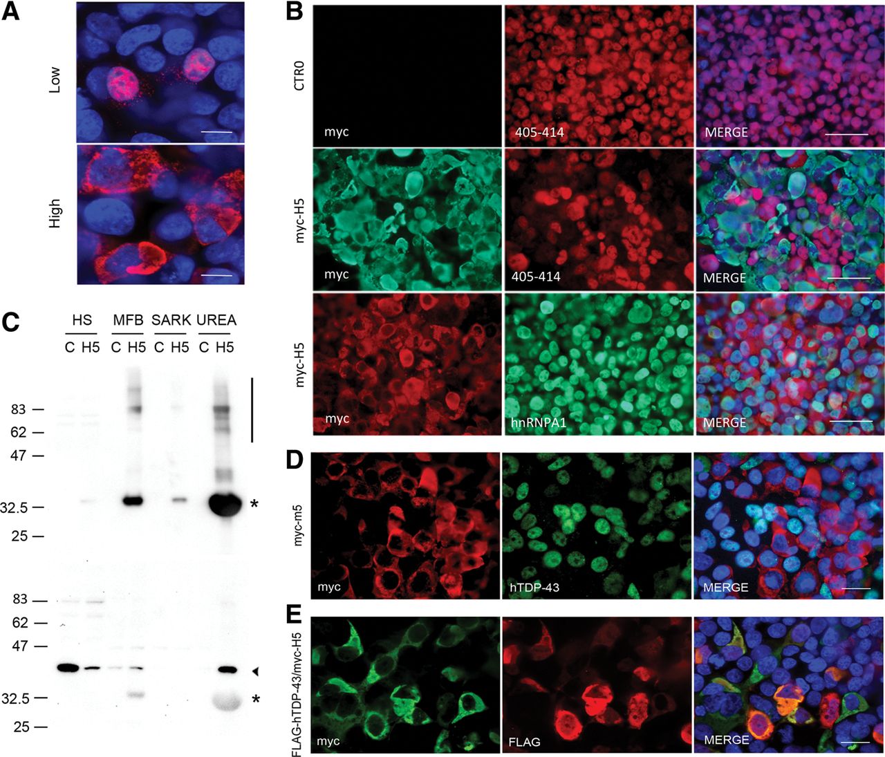
Overexpression of myc-H5 induces insolubility and loss of nuclear TDP-43. (A) Immunofluorescence of HEK293T cells transfected with myc-H5 for low expression and high expression, using myc antibody for detection. Scale bars, 10 µM. (B) Transfection with optimized protocol for high expression of empty CTR0 plasmid (top row) or myc-H5 (middle and bottom rows), followed by immunofluorescence with indicated antibodies. Note the loss of TDP-43 staining in myc-H5 cells compared with empty plasmid. Nuclear staining of another RNA-binding protein HNRNPA1 remains intact. Scale bars, 50 µm. (C) Western blotting of high salt (HS), myelin floatation buffer (MFB), sarkosyl (SARK), and urea fractions from HEK293T cells transfected with empty CTR0 plasmid control (C) or myc-H5 (H5), using myc (top) or antibody to 405–414 C-terminus of TDP-43 (bottom). Asterisk denotes myc-H5, arrowhead TDP-43. Note the high molecular weight smear indicative of protein aggregation (bar in top panel). Also note that the C-terminal antibody nonspecifically binds to myc-H5 under these conditions. (D) HEK293T cells transfected with myc-m5 and immunostained with antibodies to myc and human-specific TDP-43. Merge with DAPI counterstain on right; note cells with m5 expression lack endogenous hTDP-43 staining, both nuclear and cytoplasmic. Scale bar, 20 µm. (E) HEK293T cells cotransfected with myc-H5 and FLAG-hTDP-43, and immunostained using indicated antibodies; merge with DAPI counterstain on right. Note the colocalization of myc and FLAG epitopes in yellow. Scale bar, 20 µm.




