


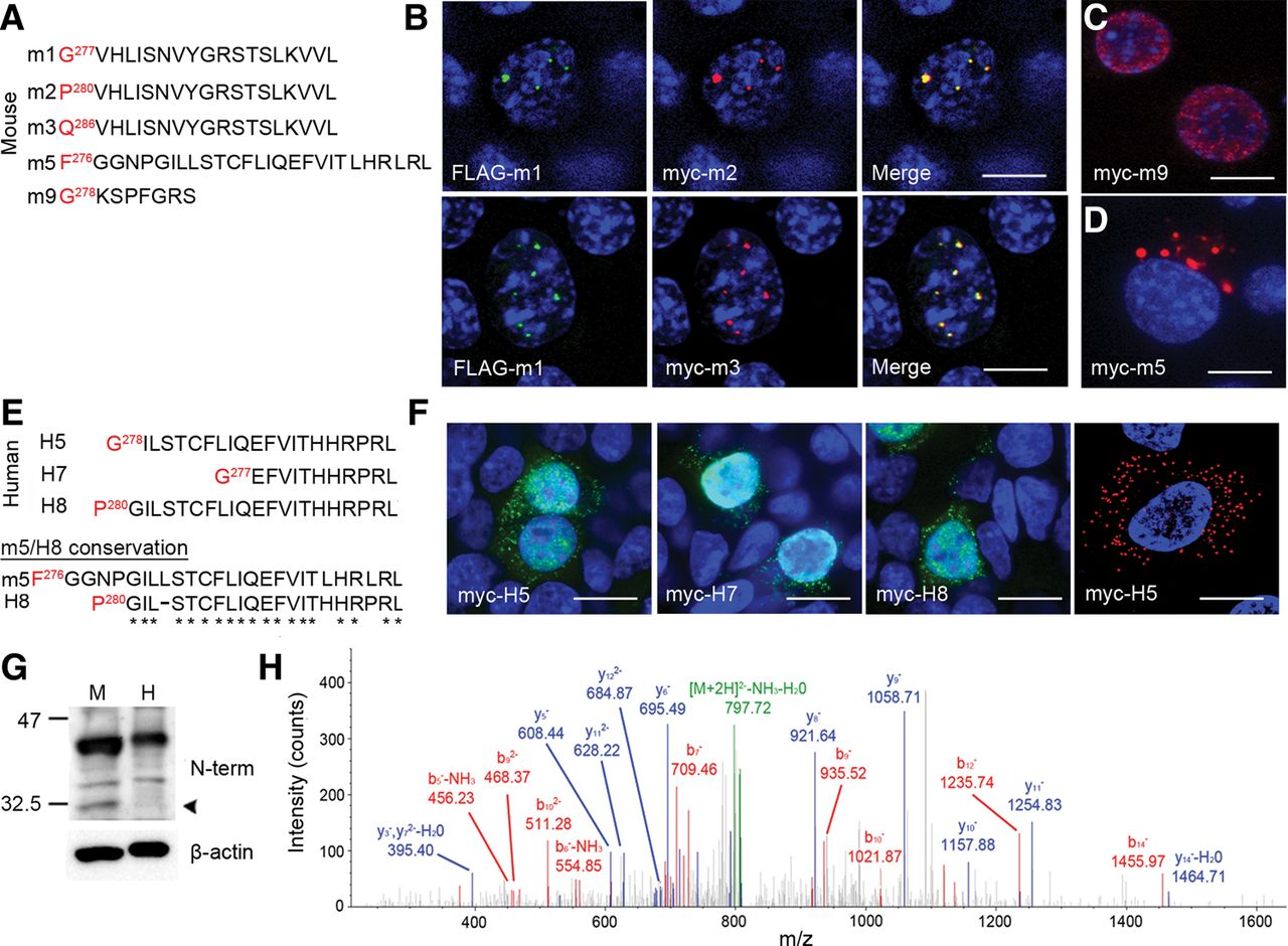
Alternative isoform expression in vitro and in vivo. (A) Alignment of sequences of murine isoforms cloned and expressed in vitro. Only the sequence after the last amino acid shared with TDP-43 (red) is shown. (B) Coexpression of FLAG-m1 and myc-m2 (top row) and FLAG-m1 and myc-m3 (bottom row) in neuro2a cells; merged images on right. (C) Expression of myc-m9 encoding an isoform with STOP codon only seven amino acids after the 6D splice junction. (D) Expression of myc-m5 in neuro2a cells. (E) Alignment of sequences of human isoforms cloned and expressed in vitro. Conservation between mouse m5 and human H8 sequence shown below. (F) Expression of myc-H5, -H7, and -H8 in HEK293T cells and myc-H5 in H4 neuroglioma cells. In B, C, D, and F, all images counterstained with DAPI; scale bars, 10 µm. (G) Western blotting of 2% SDS lysate from human and murine cortex, probed with antibody to the N-terminus of TDP-43; loading shown with β-actin. Arrowhead denotes expression of alternative isoform in murine cortex. (H) MS/MS spectra of m2 isoform peptide fragment isolated from murine cortex.




