


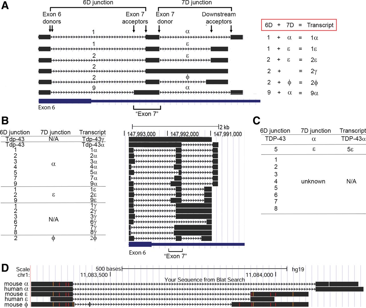
Defining a new nomenclature for Tardbp splicing. (A) Six transcripts identified in our studies (black) serve as examples for the nomenclature used in this work. Only the murine HSDR is shown. Locations of exon 6 and the region formerly known as exon 7 are indicated at bottom. Splice junctions formed between an exon 6 donor site and downstream exon 7 acceptor are termed 6D junctions and are numbered 1–9; they are exemplified here by junctions 1, 2, and 9. Note how junctions 9 and 1 share the same donor splice site, but differ in their acceptor site; however, junctions 1 and 2 differ in their donor site but share acceptor sites. Splice junctions formed between the donor exon 7 site and downstream acceptors are termed 7D junctions and are designated with the symbols α, ε, and ϕ. Combinations of 6D and 7D form specific transcripts; here these are 1α (composed of the 6D junction 1 and the 7D junction α), 1ε (6D = 1, 7D = ε), 2ε (6D = 2, 7D = ε), 2γ (6D = 2, no 7D splice junction), 2ϕ (6D = 2, 7D = ϕ), and 9α (6D = 9, 7D = α). (B) All murine transcripts identified in our studies, with 6D junction, 7D junction, and total transcript name. Position of the transcript within the HSDR is shown by alignment on the UCSC genome browser to the right of the table. (C) 6D and 7D junctions, and complete transcripts identified by RT-PCR from human neuroglioma cells. Owing to primer position, we only detected one “pairing” of 6D and 7D, hence the 7D junctions that might pair with 6D junctions are majority “unknown.” (D) Conservation between mouse and human 7D splice junctions. Sequences shown are aligned on the human UCSC genome browser. Mismatched base pairs appear as orange or red.




