


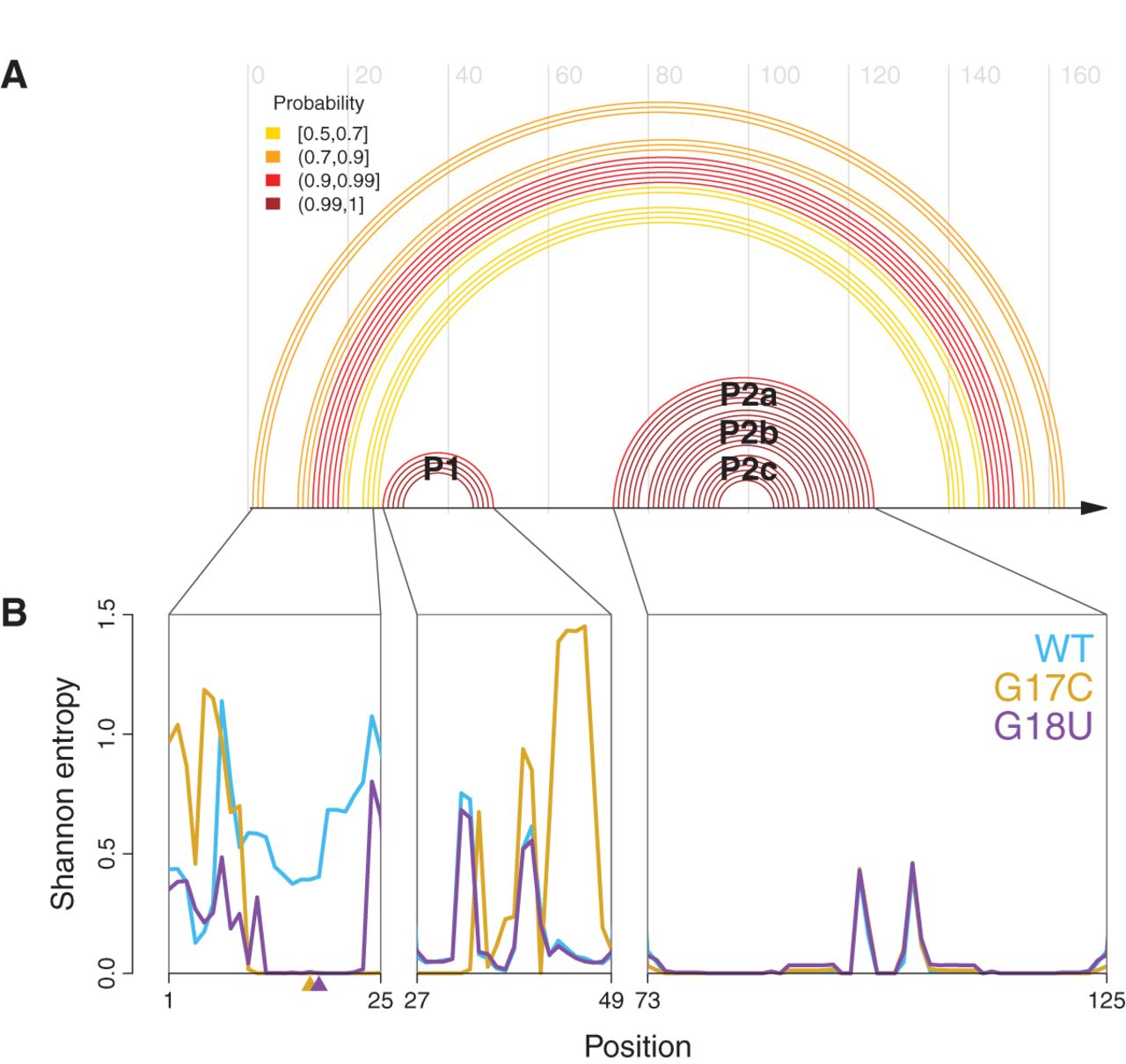
Elements of the core structure in the 5′ UTR can be disrupted by mutation. (A) WT base-pair probabilities >50% computed using a SHAPE-directed free energy function (Deigan et al. 2009; Wilkinson et al. 2009), plotted as an arc diagram; they reveal a core structure containing two helices, P1 and P2. The P2 helix has two bulges yielding the P2a, P2b, and P2c paired regions. Peripheral helices are lower probability and thus more variable. (B) Shannon entropies for individual nucleotides in WT (blue), G17C (gold), and G18U (purple). The two retinoblastoma-associated mutations drastically reduce the entropy of nucleotides 13–23 relative to WT, and G17C changes the pattern of entropy over the P1 helix. The P2 helix has low entropy in all three constructs, indicating a well-defined structure and suggesting the mutations do not alter this region of the mRNA.




