


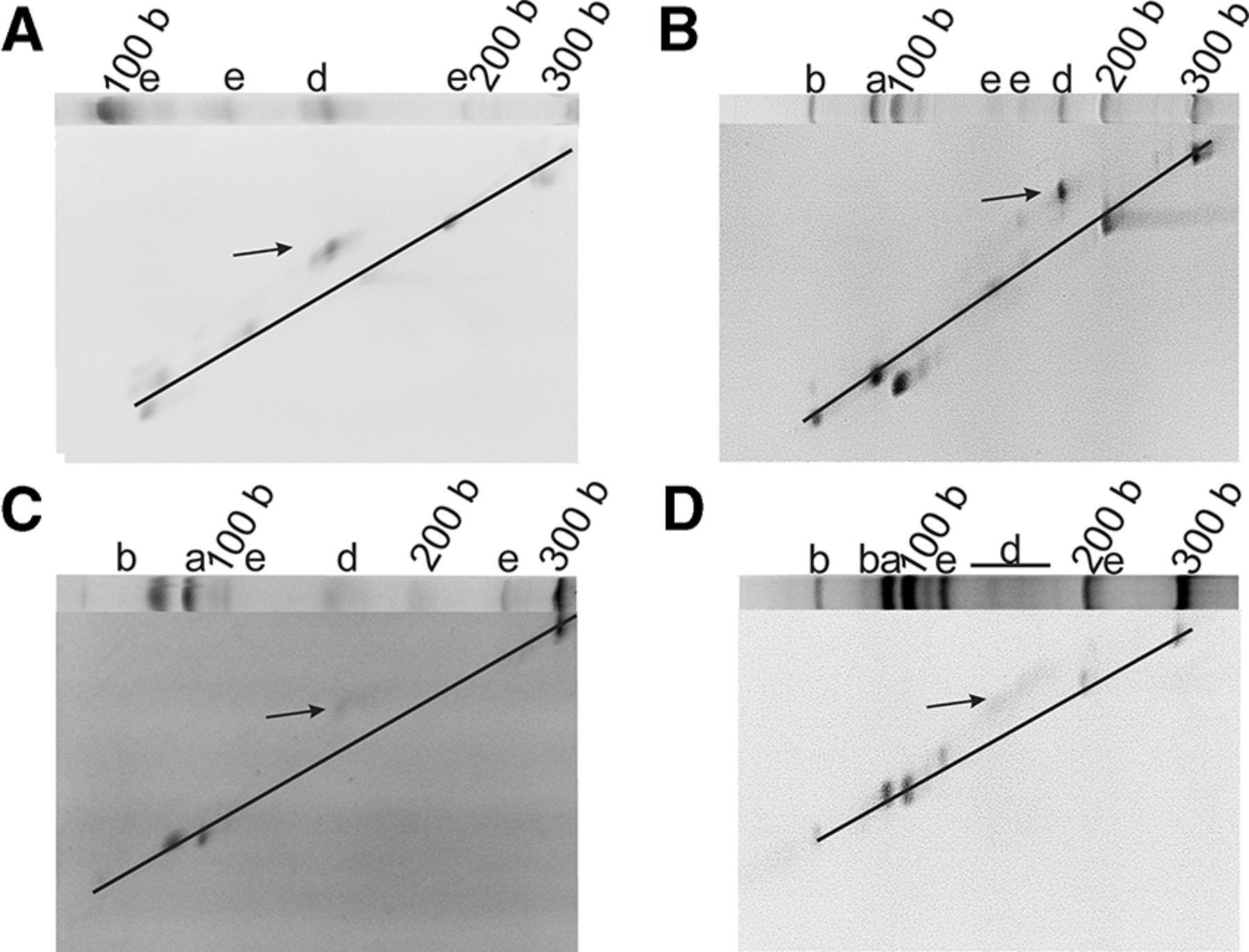
FIGURE 3.
Two-dimensional gel electrophoretic analysis of PBD1 (A), PBD2 (B), PBD3 (C), and PBD4 (D). All samples were mixed with a linear RNA size standard prior to subjecting onto the gel. The first dimension gel of the respective system is implemented in each panel. The diagonal marks the linear RNAs; circular species are denoted by an arrow.




