


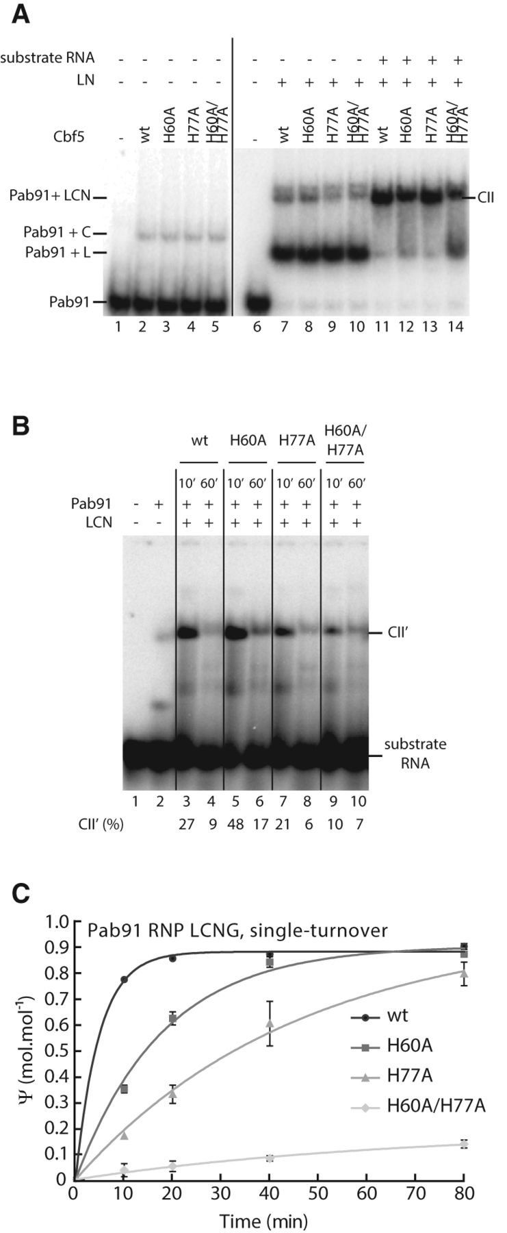
Contribution of both histidines H60 and H77 for the guide RNA-dependent activity of Cbf5 within the Pab91 RNP. (A) Analysis by electrophoretic mobility shift assay (EMSA) of various box H/ACA sub-sRNPs and the complex formed between the box H/ACA sRNP and a substrate RNA. The guide RNA Pab91 which was radiolabeled at the 5′ end with 32P was incubated with the wild type (wt) or the variant forms of Cbf5 (variants H60A, H77A, and H60A/H77A) as indicated above each lane, alone (lanes 2–5) or in the presence of L7Ae and Nop10 (lanes 7–14) and in the absence (lanes 7–10) or the presence of the substrate RNA 22-U (lanes 11–14). After 10 min incubation at 65°C, the different complexes were fractionated by nondenaturing polyacrylamide gel electrophoresis. The identity of the sub-RNPs is indicated: C and LCN correspond, respectively, to the Pab91–Cbf5 and the Pab91–L7Ae–Cbf5–Nop10 complexes; the CII complex is formed upon association of the Pab91 LCN RNP and the substrate RNA (Charpentier et al. 2005). (B) Analysis by EMSA of the complex formed between the RNP enzyme and the substrate RNA (complex CII′). The substrate RNA 22-U which was radiolabeled at the 5′ end with 32P was incubated with the wild-type and variant Pab91 LCN RNP assembled with the unlabeled Pab91 sRNA and the L7Ae, Cbf5, and Nop10 proteins as described in Materials and Methods. After 10 and 60 min at 65°C, the different complexes were fractionated by nondenaturing polyacrylamide gel electrophoresis. (C) Time course analysis of Ψ formation by the Pab91 LCNG RNP enzyme in the 22-U substrate RNA. The uniformly [α-32P] CTP radiolabeled substrate RNA 22-U was incubated with the guide RNA Pab91 and proteins LCNG wild-type (in black), H60A (in dark gray), H77A (in gray), and H60A/H77A (in light gray) and analyzed by 1D-TLC as described in Materials and Methods. The radioactivity was quantified by PhosphorImager analysis. The quantities of Ψ nucleotides formed are expressed in moles per mole of 22-U.




