


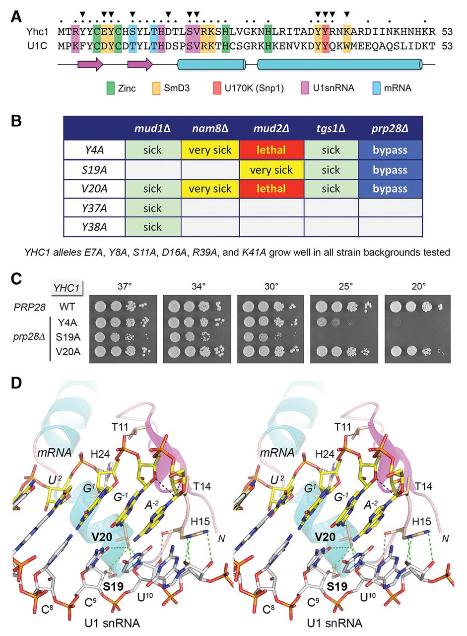
Structure-guided mutational analysis of Yhc1. (A) Alignment of the primary structures of the N-terminal domains of S. cerevisiae Yhc1 and human U1-C. Positions of side-chain identity/similarity are indicated by • above the alignment. The secondary structure elements are depicted below the alignment, with β strands as magenta arrows and α helices as cyan cylinders. U1-C/Yhc1 amino acids that coordinate zinc, make contacts to other U1 snRNP subunits, and contact the U1 snRNA or the mRNA 5′ splice site sequence are highlighted in color-coded boxes as indicated. (B) Summary of genetic interactions of the indicated YHC1-Ala alleles. (C) Prp28 bypass. Yeast prp28▵ yhc1▵ cells harboring the indicated YHC1 allele on a CEN LEU2 plasmid and either wild-type PRP28 (CEN HIS3) or an empty CEN HIS3 plasmid (prp28▵) were spot-tested for growth on YPD agar at the temperatures specified. (D) Stereo view of the human U1 snRNP structure highlighting the interactions of U1-C/Yhc1 (depicted as a cartoon trace with magenta β strands and cyan helices, with selected amino acids as stick models with beige carbons) with the RNA duplex formed by the U1 snRNA 5′ leader (5′-ACUUAC8C9U10, depicted as a stick model with gray carbons) and the mRNA 5′SS exon–intron junction (5′-A−2G−1/G1U2, depicted as a stick model with yellow carbons). Protein–RNA contacts are indicated by dashed lines: black for hydrogen bonds and green for van der Waals interactions.




