


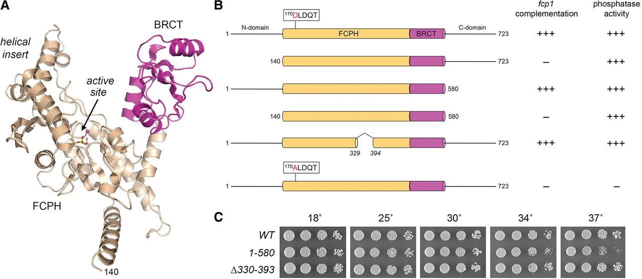
Requirements for SpFcp1 activity in vivo. (A) Tertiary structure of SpFcp1(140–580) from pdb 3EF1. The core catalytic FCPH module sits at the base of the Y-shaped protein. The left prong of the Y is formed by a helical module inserted into the FCPH domain; the right prong is a BRCT domain (magenta). The active site (arrow) is demarcated by a Mg2+ ion (magenta sphere) and Asp170-BeF3 adduct (stick model). (B) Cartoon representations of the wild-type and mutated Fcp1 proteins. The FCPH and BRCT modules within the central catalytic domain are denoted as beige and magenta cylinders. The position of the signature DxDxT motif of the acyl-phosphatase superfamily is highlighted. The flanking N and C domains are horizontal lines. The column at right indicates whether the respective recombinant Fcp1 proteins have phosphatase activity in vitro. The column at left indicates whether the fcp1 alleles encoding the indicated proteins are functional in vivo in S. pombe. Lethal alleles are indicated by −. (C) Exponentially growing cultures of S. pombe strains with the indicated chromosomal fcp1 alleles were adjusted to A600 of 0.1 and aliquots of serial fivefold dilutions were spotted to YES agar and incubated at the indicated temperatures.




