


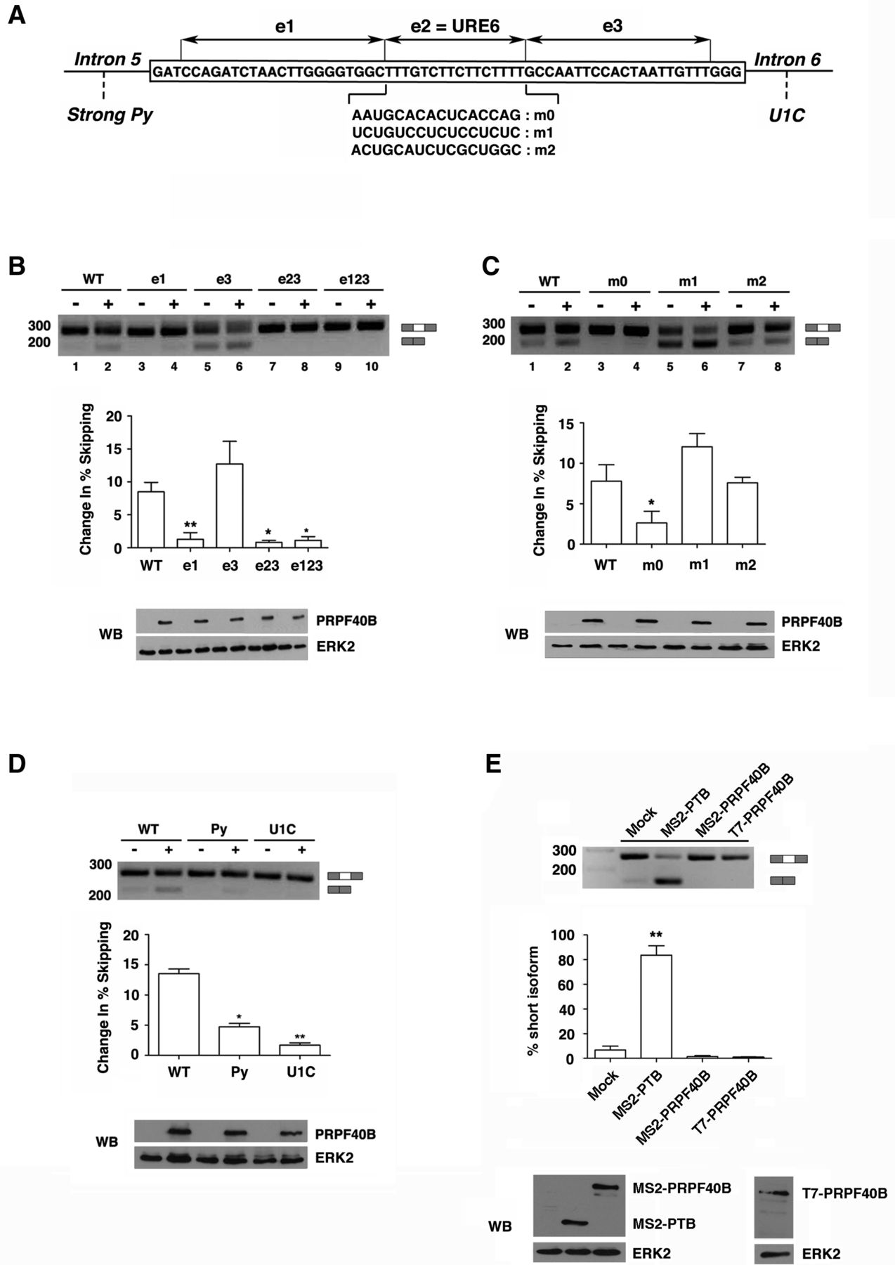
Exon 6 sequences and associated 5′ and 3′ splice sites are required for PRPF40B-mediated Fas splicing regulation. (A) Schematic representation of the structure of the Fas gene, including the genomic sequence of exon 6 in the box and the neighboring introns 5 and 6. e1, e2, and e3 represent three segments in exon 6, where e2 corresponds to the uridine-rich exonic silencer (URE6). m0, m1, and m2 represent different RNA sequences replacing the URE6 silencer. The strong Py in intron 5 indicates the substitution of a weak 3′ splice site associated with exon 6 with a strong polypyrimidine tract. The U1C in intron 6 corresponds to a mutant strengthening the 5′ splice site by improving its base-pairing potential with U1 snRNA. (B–D) Analyses of the effects of Fas splice site selection in response to PRPF40B overexpression. HEK293T cells were cotransfected together with the corresponding Fas minigene and a plasmid expressing PRPF40B. RT-PCR was performed to analyze the alternatively spliced forms of the Fas minigenes. The graphs show the densitometric analysis results as the change in exon skipping average from three independent experiments (means ± SEM). (*) P < 0.05; (**) P < 0.01. Cell lysates were analyzed by immunoblotting with the indicated antibodies to detect the PRPF40B and ERK2 proteins. (E) Effect of tethering PTB and PRPF40B to exon 6 through the RNA MS2-binding domains. The sequence of the URE6 element was replaced by two MS2 stem–loop binding sites in tandem. This minigene was cotransfected with plasmids expressing the indicated proteins or empty vector. RT-PCR was performed to assess the alternatively spliced isoforms of Fas. The bar graph shows the densitometric analysis results as the exon skipping average from three independent experiments (means ± SEM). (**) P < 0.01. A fraction of the cell lysates was analyzed by immunoblotting to detect the indicated proteins.




