


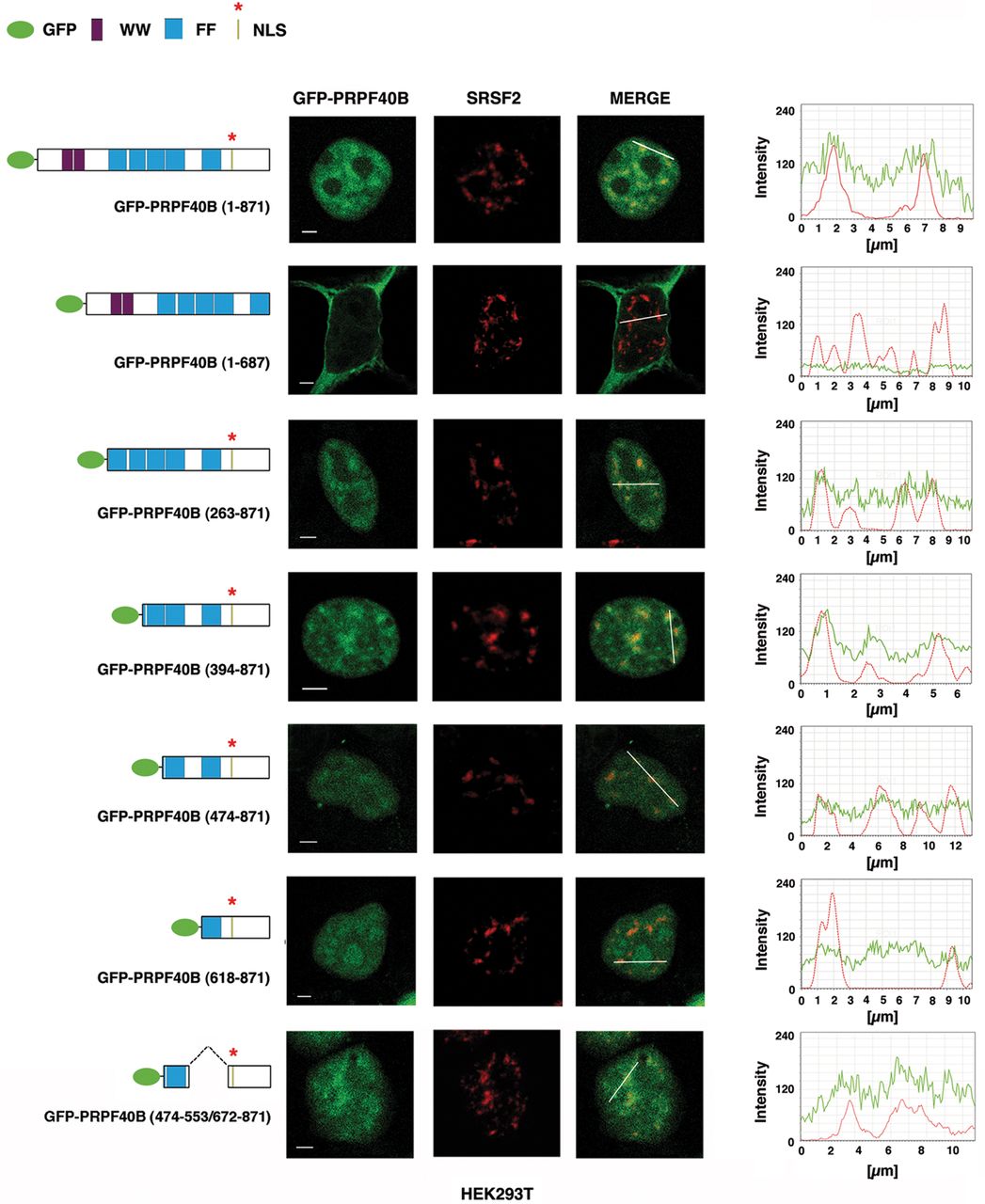
Subcellular localization of PRPF40B. HEK293T cells expressing EGFP-tagged PRPF40B constructs (green) were immunolabeled with SRSF2 antibody (red). Schematic diagrams of the EGFP-PRPF40B fusion proteins are shown at the left of each panel. The numbers in parentheses represent the PRPF40B amino acids contained in the construct. GFP protein, the two WW domains, the putative nuclear localization signal (NLS), and the five FF domains are indicated. Individual and merged images of the cell are shown. Line scans showing the local intensity distribution of the dual staining are represented to the right of the panels. The bars in the merged panels indicate the position of the line scans. Bar, 3 mm.




