


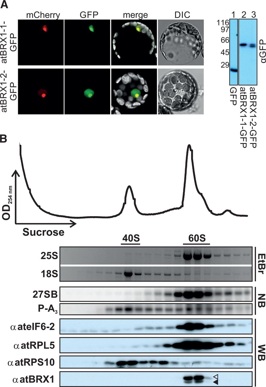
Both atBRX1 proteins are nucleolar and associated with pre-60S complexes. (A) A. thaliana protoplasts were cotransformed with plasmids coding for Fibrillarin-mCherry and either atBRX1-1-GFP (top) or atBRX1-2-GFP (bottom). mCherry fluorescence (left) and GFP fluorescence (second panel) were merged with the autofluorescence of chlorophyll (third panel). A differential interference contrast (DIC) image is also shown (fourth panel). A representative image is given. Note that the nuclear signals may partially be due to overexpression of fusion proteins from the 35S promoter. (Right) Transformed protoplasts were lysed and subjected to Western blot using a GFP antibody. (B) Sucrose density gradient centrifugation using total cell lysate from A. thaliana cell culture was performed. The UV absorption profile is shown on top. Mature rRNAs in the fractions were visualized by ethidium bromide (EtBr) staining. Pre-rRNAs were detected by Northern blot (NB). Western blot (WB) analysis was carried out using the antibodies indicated on the left. Arrowheads indicate the migration of atBRX1-1 (filled) and atBRX1-2 (open).




