


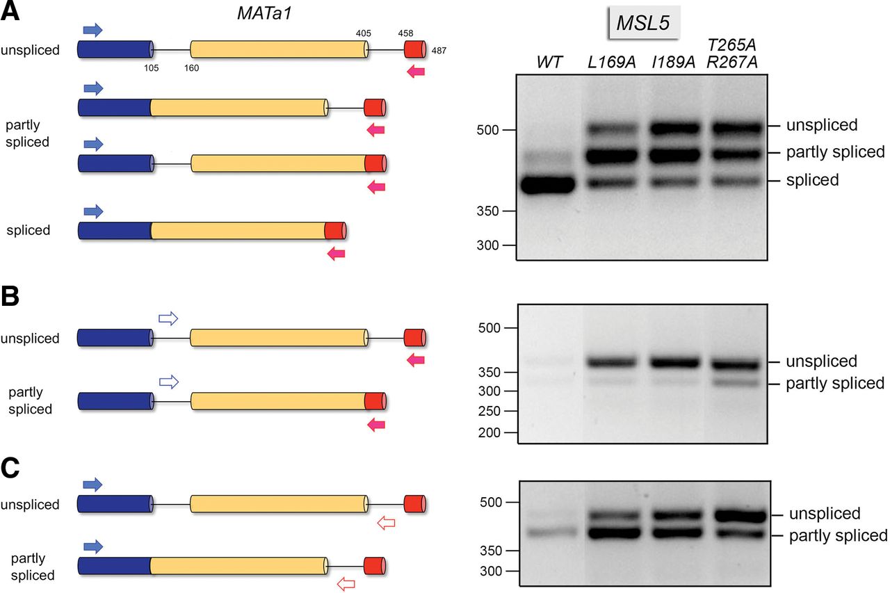
Effects of MSL5 mutations on MATa1 pre-mRNA splicing. The unspliced MATa1 pre-mRNA comprising three exons (depicted as horizontal cylinders with exon 1 in blue, exon 2 in gold, and exon 3 in red) and two introns, the two partly spliced intermediates with one intron retained, and the mature spliced mRNA are depicted on the left. RNA isolated from MSL5 wild-type (WT), L169A, I189A, and T265A-R267A cells was reverse transcribed with an oligo(dT) primer, and the MATa1 cDNAs were PCR-amplified with gene-specific primer pairs, depicted as forward and reverse arrows, in exon 1 and exon 3 (A), intron 1 and exon 3 (B), or exon 1 and intron 2 (C). The PCR products were resolved by native agarose gel electrophoresis and visualized by staining with ethidium bromide. All lanes in the image are from the same gel and photographic exposure, from which intervening lanes were cropped and the WT lane was moved laterally to be next to L169A; the T265A-R267A lane was moved laterally to be next to I189A. The positions and sizes (bp) of linear duplex DNA markers are indicated on the left. The positions of the RT-PCR products of unspliced, partly spliced, and fully spliced MATa1 are indicated at right.




