


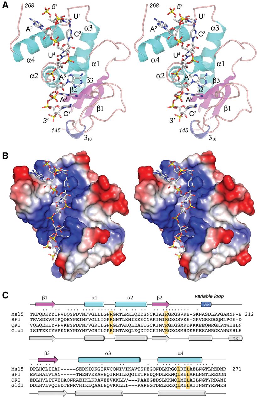
Structure of the Msl5-(KH-QUA2)·branchpoint RNA complex. (A) A stereo view of the tertiary structure is shown as a cartoon model, with β strands colored magenta, α helices colored cyan, and a 310 helix colored blue. The N and C termini (amino acids 145 and 268) are indicated. The branchpoint RNA is depicted as a stick model. (B) A stereo view of a surface electrostatic model of Msl5-(KH-QUA2), generated in Pymol, is shown, with the branchpoint RNA depicted as a stick model. (C) The secondary structure elements of yeast Msl5-(KH-QUA2) (colored as in A) are shown above the Msl5 amino acid sequence, with β strands rendered as arrows and helices as cylinders. The Msl5 primary structure is aligned to those of the homologous human SF1, human QKI, and C. elegans GLD-1 proteins. Gaps in the alignment are indicated by dashes. Positions of amino acid side chain identity/similarity in all four polypeptides are indicated by dots above the sequence. The secondary structure elements of GLD-1 (colored gray) are shown below its amino acid sequence. The four conserved amino acids that are important per se for Msl5 function in vivo (as determined previously by alanine scanning) are highlighted in yellow boxes.




