


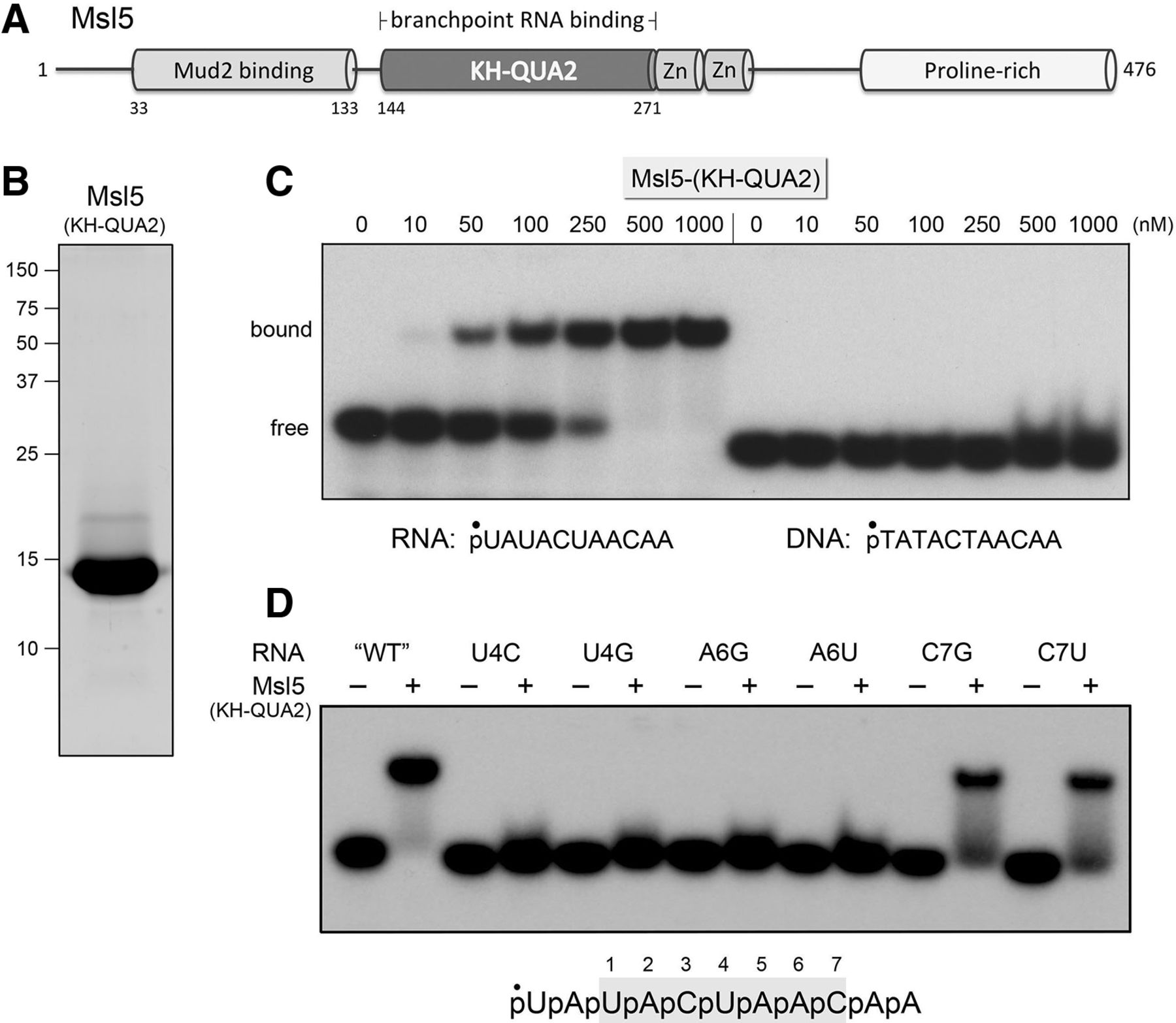
Msl5-(KH-QUA2) is an autonomous branchpoint-specific RNA binding domain. (A) Domain organization of yeast Msl5. The 476-amino acid Msl5 polypeptide is depicted as a linear array with the N-terminus at left and the C-terminus at right, and the known or imputed domains drawn as cylinders spanning their segments of the primary structure. (B) An aliquot (7.5 µg) of purified recombinant Msl5-(KH-QUA2) (amino acids 147–271) was analyzed by SDS-PAGE. The Coomassie blue-stained gel is shown. The positions and sizes (kDa) of marker polypeptides are indicated at left. (C) Nucleic acid binding reaction mixtures (10 µL) containing 25 mM Tris–HCl, pH 7.5, 1 mM DTT, 10% glycerol, 100 nM 5′ 32P-labeled 11-mer RNA (left panel) or DNA (right panel) oligonucleotide as specified, and Msl5-(KH-QUA2) as specified were incubated at 4°C for 20 min. The mixtures were analyzed by electrophoresis at 4°C for 90 min at 110 V through a 15-cm native 10% polyacrylamide gel containing 22.5 mM Tris–borate, 2.5% glycerol. An autoradiograph of the gel is shown. The species corresponding to free and Msl5(KH-QUA2)-bound oligonucleotides are indicated at left. (D) RNA binding reaction mixtures containing 25 mM Tris–HCl, pH 7.5, 1 mM DTT, 10% glycerol, 100 nM 5′ 32P-labeled 11-mer RNA as specified, and either no protein (−) or 500 nM Msl5-(KH-QUA2) (+) were incubated at 4°C for 20 min. The mixtures were analyzed by native PAGE as in C. An autoradiograph of the gel is shown. The wild-type branchpoint-containing RNA is shown at bottom, with the consensus yeast branchpoint sequence shaded gray and the seven branchpoint nucleobases numbered from 5′ to 3′.




