


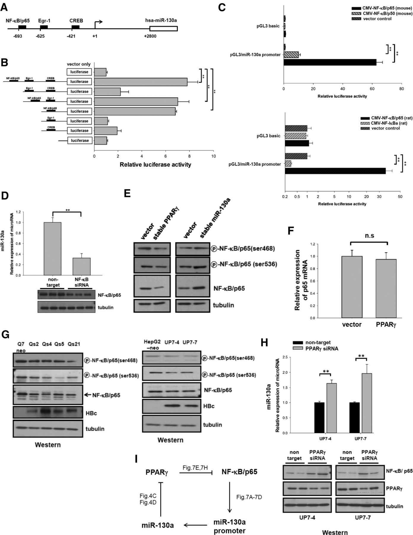
A feed-forward loop between NF-κB/p65 and miR-130a via PPARγ. The promoter activity of miR-130a can be stimulated by NF-κB/p65, while PPARγ appeared to reduce the protein level of NF-κB/p65. (A) A schematic diagram shows several potential binding sites for transcription factors in the putative hsa-miR-130a promoter element upstream of the transcription start site (+1): A putative NF-κB/p65 binding site at −693 position was as previously reported (Zhou et al. 2010a), and the sites at −625 and −421 for Egr-1 and CREB were predicted using the online softwares Promo and TRANSFAC (Wingender et al. 2000; Messeguer et al. 2002). (B) Analysis of miR-130a promoter by deletion mapping and reporter assay. Briefly, HuH-7 cells were transfected with various luciferase reporter constructs containing sequentially deleted hsa-miR-130a promoter. Deletion of an NF-κB/p65 binding site resulted in significantly reduced reporter activity. (C) (Upper panel) Both NF-κB/p65 and p50 can stimulate miR-130a promoter activities by ∼60-fold and 10-fold, respectively. HuH-7 cells were cotransfected with expression vectors of either pCMV-mouse NF-κB/p65 or p50, and a luciferase reporter driven by an hsa-miR-130a promoter. (Lower panel) A 30-fold stimulation effect on miR-130a promoter was obtained by using a rat pCMV-flag- NF-κB/p65. Cotransfection with a rat IκB expression vector repressed the luciferase activity by about fourfold. Relative luciferase values for each transfection were normalized with cotransfected renilla expression. (D) Conversely, knockdown of the endogenous NF-κB/p65 by transfection with siRNA resulted in decreased expression of miR-130a by stem–loop qPCR analysis. (E) (Left panel) A PPARγ stable expression cell line of HepG2 origin exhibited reduced levels of phosphorylated and total NF-κB/p65 protein by Western blot analysis. (Right panel) In contrast, a miR-130a stable expression cell line exhibited increased levels of phosphorylated and total NF-κB/p65 protein. (F) The relative mRNA level of NF-κB/p65 was not affected in stable PPARγ expressing HepG2 cells by qPCR. (G) The reduction of phosphorylated and total NF-κB/p65 protein was observed in stable HBV-producing rat (left panel) or human (right panel) hepatoma cells by Western blot analysis. (H) Knockdown of the endogenous PPARγ by siRNA treatment was conducted in stable HBV-producing human hepatoma cells UP7-4 and UP7-7. This experiment resulted in increased expression of miR-130a by stem–loop qPCR (upper panel) and NF-κB protein production by Western blot analysis (lower panel). (I) A cartoon summarizes a positive feed-forward loop between miR-130a and NF-κB/p65 via PPARγ. (**) P < 0.05.




