


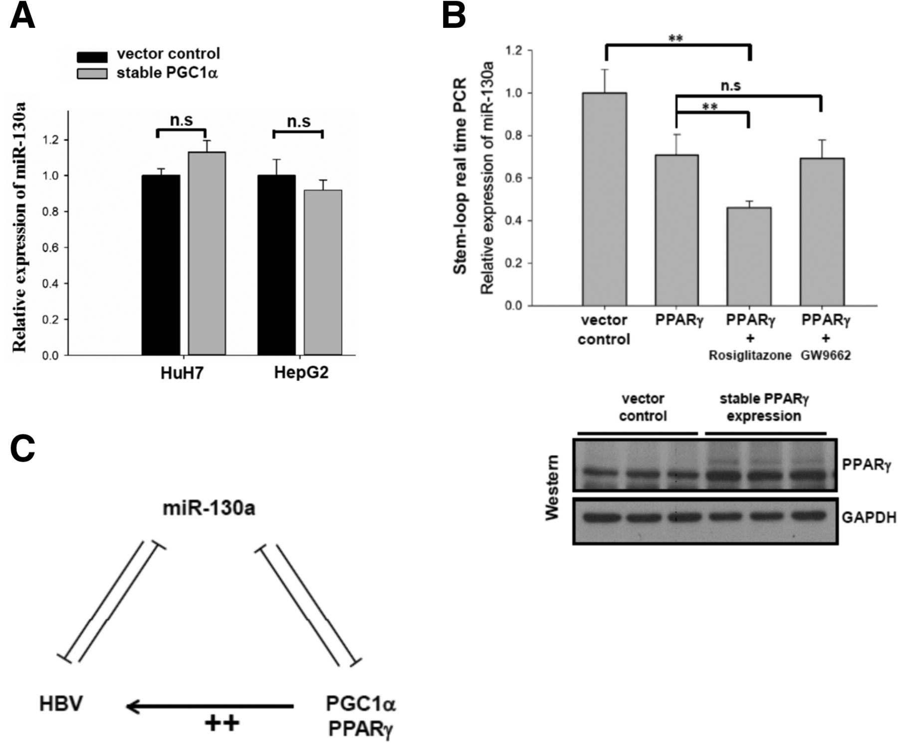
The effect of PGC1α and PPARγ on the expression of miR-130a. (A) The expression level of miR-130a was not affected in stable PGC1α-expressing HepG2 or HuH7 cell lines. (B) (Upper panel) Reduction of miR-130a was observed in PPARγ-expressing HepG2 cell lines using stem–loop qPCR. U6 snRNA was used as an internal control for normalization. Rosiglitazone, but not GW9662, further reduced the expression of miR-130a. (Lower panel) Increased amounts of PPARγ protein in stable PPARγ-expressing HepG2 cell lines were detected by Western blot. (C) This cartoon summarizes the relationships among PPARγ, PGC1α, miR-130a, and HBV. A feed-forward amplification loop among HBV, PGC1α, and PPARγ can be mediated through a miR-130a intermediate. The symbol ++ represents an enhanced stimulatory effect from the combination of PGC1α and PPARγ.




