


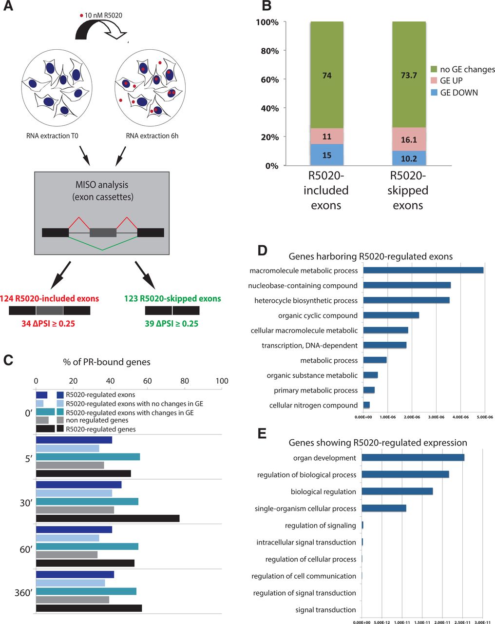
Progesterone-induced alternative splicing changes in T47D breast cancer cells. (A) Scheme summarizing the experimental procedure and number of alternative splicing changes identified. (B) Fraction of alternative exon changes (increased inclusion or skipping) that occurs in genes without expression changes (no change), up-regulated (up), or down-regulated (down) genes. (C) Fraction of expressed genes at different times after hormone treatment that contain progesterone receptor (PR) binding sites. Different gene categories are represented with different colors, as indicated. (D,E). Gene ontology terms enrichment for progesterone-regulated exons (D) and genes (E). Statistical significance (P-value) of GO terms enrichment is indicated.




