


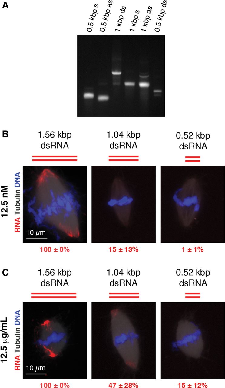
Spindle localization of dsRNAs of different lengths. (A) Native agarose gel of keratin-19 RNA constructs used in the experiments in B. Gel is stained with ethidium bromide. (B) Fluorescence imaging of RNA localization to spindles using dsRNAs of different lengths that do not contain single-stranded overhangs. Equivalent moles of RNA were added to each sample for a final RNA concentration of 12.5 nM. RNA (red) is labeled with Cy3-CTP, DNA (blue) is stained with DAPI, and microtubules (white) are visualized by the addition of HiLyte Fluor 488-tubulin. Scale bar is 10 μm. The average percentage of spindles exhibiting localized RNA from three biological replicates is reported in red text. (C) Same as in B, except that an equivalent mass of RNA was added to each sample for a final RNA concentration of 12.5 μg/mL.




