


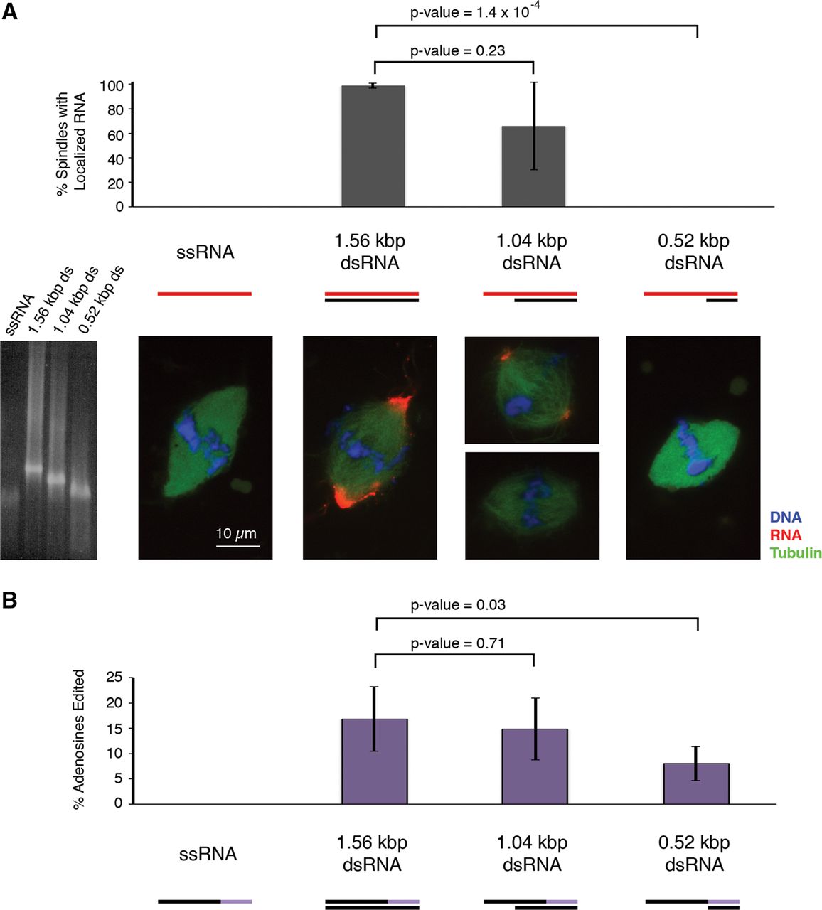
Effect of dsRNA length on spindle localization and editing. (A) Quantification of the percentage of spindles exhibiting localized RNA in samples containing keratin-19 RNA constructs with various lengths of double-stranded structure. Data are the average of three biological replicates. P-values were calculated using a two-sided paired t-test. Below the bar graph are representative images of the spindles for each RNA construct. RNA (red) is labeled with Cy3-CTP, DNA (blue) is stained with DAPI, and microtubules (green) are visualized by the addition of HiLyte Fluor 488-tubulin. Scale bar is 10 μm. A native agarose gel of the RNA constructs stained with ethidium bromide is shown. (B) Quantification of the percentage of adenosines edited in the RNA constructs during the experiment in A. Editing was assessed in the terminal third of the sense strand (depicted in purple). P-values were calculated using a two-sided Welch's t-test.




