


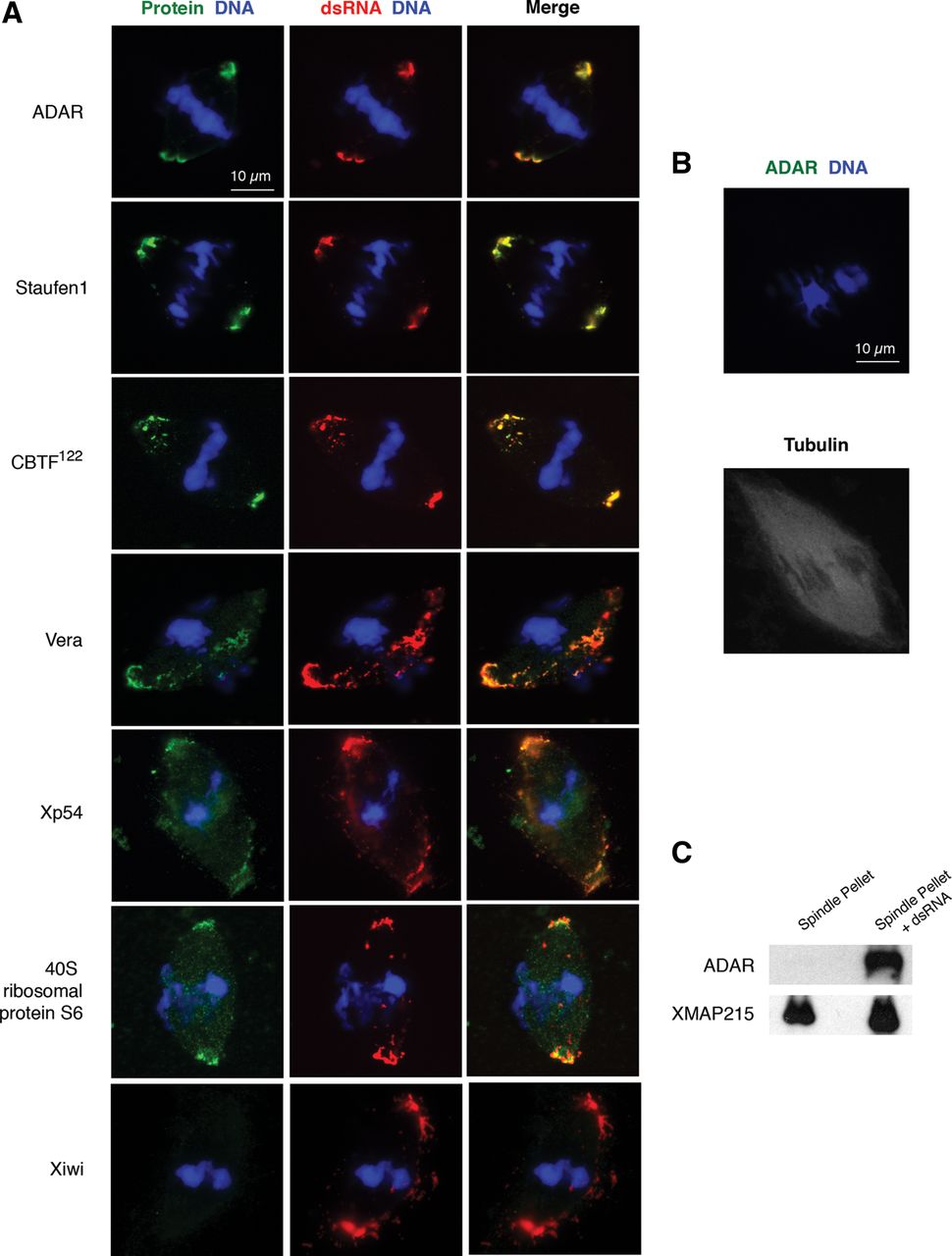
Characterization of the spindle-localized dsRNA complex. (A) Immunofluorescence imaging of candidate RNA-interacting proteins (green) on spindles with localized, exogenous dsRNA (red). Double-stranded keratin-19 RNA is labeled with Cy3-CTP, DNA (blue) is stained with DAPI, and secondary antibodies are conjugated to Alexa 488. Scale bar is 10 μm. (B) ADAR staining (green) on spindles in the absence of exogenous dsRNA. DNA (blue) is stained with DAPI, secondary antibody is conjugated to Alexa 488, and microtubules (second panel, white) are visualized by the addition of rhodamine-tubulin. Scale bar is 10 μm. (C) Western blots on pelleted spindles isolated from egg extract with or without exogenous double-stranded kertain-19 RNA. XMAP215, a microtubule-binding protein, serves as a loading control.




