


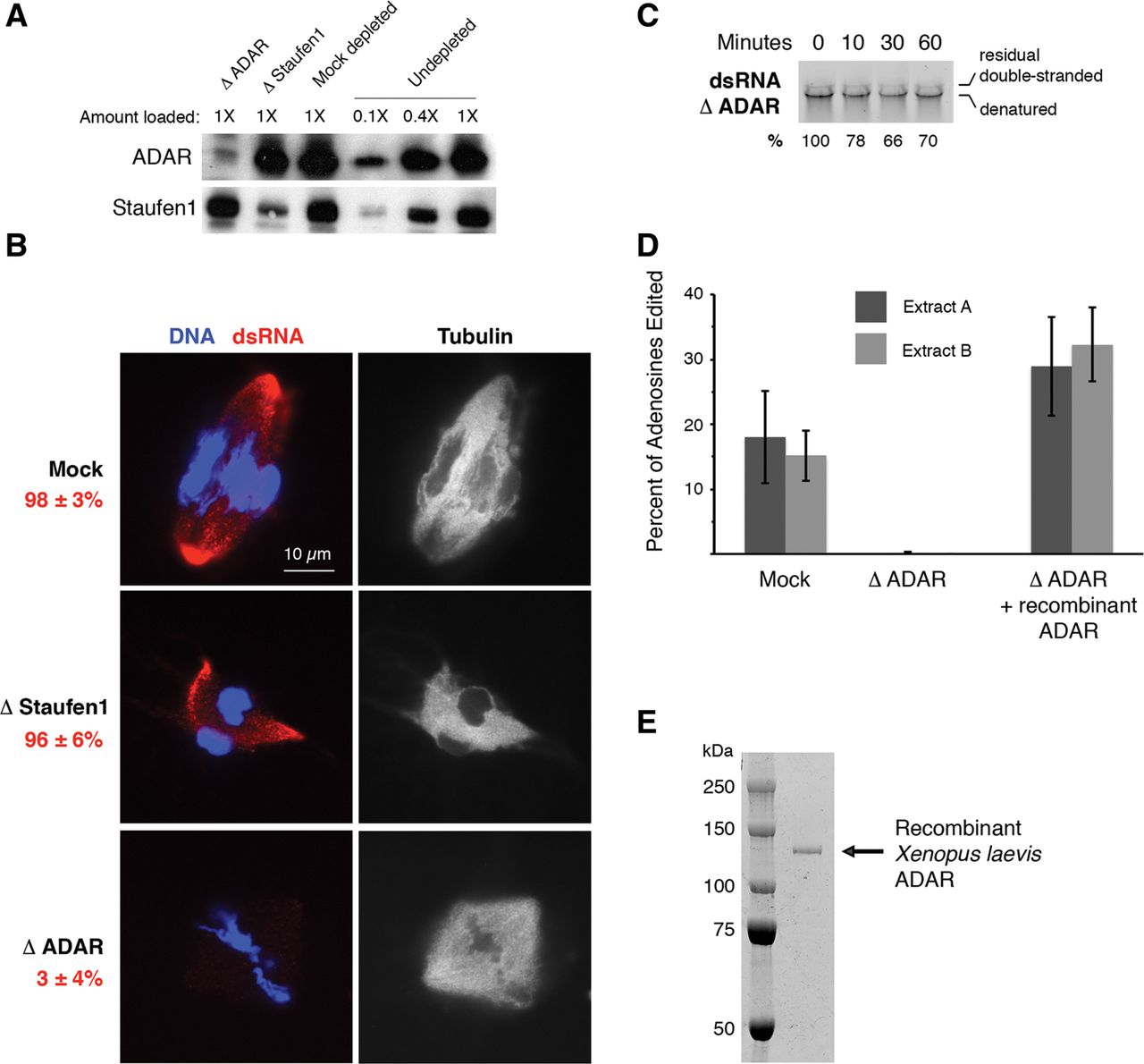
Requirement of an ADAR-containing complex for the spindle localization of dsRNA. (A) Western blot demonstrating immunodepletion of ∼90% of ADAR and ∼75% of Staufen1 from egg extract. (B) Fluorescence imaging of dsRNA localization to spindle poles in mock, Staufen1, or ADAR-depleted egg extract. The average percentage of spindles exhibiting localized RNA from three biological replicates is reported in red text. Double-stranded keratin-19 RNA (red) is labeled with Cy3-CTP, DNA (blue) is stained with DAPI, and microtubules (white) are visualized by the addition of HiLyte Fluor 488-tubulin. Scale bar is 10 μm. (C) Time course of dsRNA stability in ADAR-depleted egg extract. Percent of double-stranded keratin-19 RNA remaining is relative to 0 min. (D) Quantification of the average percentage of adenosines edited within a 300 base region on one strand of double-stranded IME1 RNA incubated for 40 min in egg extract. Experiment was performed in mock-depleted egg extract, ADAR-depleted egg extract, and ADAR-depleted egg extract rescued with 1 μM purified recombinant ADAR. (E) Coomassie-stained polyacrylamide gel of purified recombinant X. laevis ADAR.




