


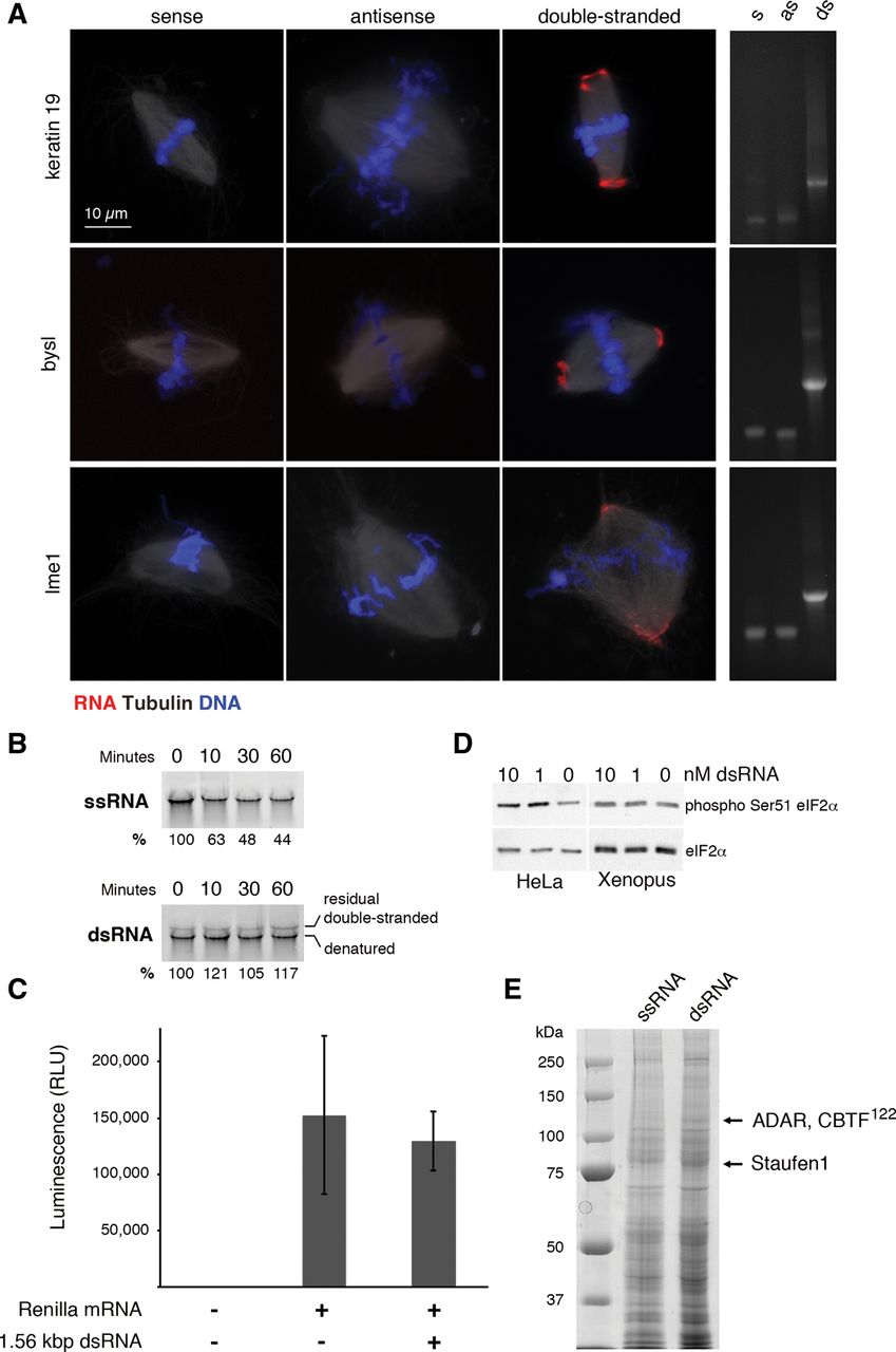
Fate of exogenous dsRNA added to Xenopus egg extract. (A) Fluorescence imaging of Cy3-labeled RNA (red) added to egg-extract spindles at a final concentration of 12.5 nM. DNA (blue) is stained with DAPI, and microtubules (white) are visualized by the addition of HiLyte Fluor 488-tubulin. Single-stranded RNAs correspond to the sense or antisense strand of a 1.56 kb fragment from the X. laevis keratin-19 cDNA, a 1.75 kb fragment from the X. laevis bysl cDNA, or a 1.5 kb fragment from the Saccharomyces cerevisiae IME1 promotor. Double-stranded RNAs were formed by annealing the sense and antisense transcripts prior to addition to egg extract. For all three dsRNAs, the average percentage of spindles exhibiting RNA-localization from three biological replicates was >90%. Scale bar is 10 μm. Native agarose gels of the sense (s), antisense (as), and double-stranded (ds) RNAs stained with ethidium bromide are presented to the right of the images. (B) Time course of the stability of single- and double-stranded keratin-19 RNA in egg extract. Percent of RNA remaining is relative to 0 min. (C) Luciferase assay performed in egg extract with or without 1.56 kbp double-stranded keratin-19 RNA added at 12.5 nM. (D) Western blots for eIF2α and phosphorylated eIF2α in HeLa cell extract and Xenopus egg extract with or without added dsRNA. (E) SDS-PAGE of egg-extract proteins that coprecipitate with exogenous single- or double-stranded keratin-19 RNA coupled to magnetic beads. Arrows indicate two bands that reproducibly coprecipitate only with dsRNA. The most abundant components of these bands, as determined by mass spectrometry, are listed.




