


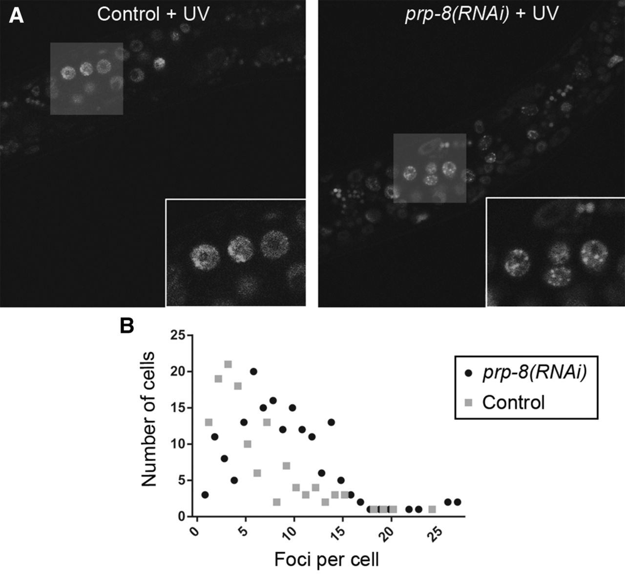
Higher sensitivity to UV damage observed in prp-8(RNAi)-treated animals. After UV exposure, foci formation evidenced by the transgene opIs263 [Prpa-1::rpa-1::YFP+unc-119(+)] is more abundant in animals with partial inactivation of prp-8 compared with control worms (fed with empty vector clone). (A) Representative confocal images of transgenic worms carrying the RPA-1::YFP transgene under control and prp-8(RNAi) conditions. RNAi treatment started from L1 stage at 20°C. After 24 h, animals were exposed to a 100 J/m2 dose of UV-C radiation, and 24 h later foci formation was assessed through confocal microscopy. Foci formation was observed in somatic and germline cells in both conditions. White squares show magnified images of germline cells with RPA-1::YFP foci. (B) Quantification of foci formation per cell represented in a dispersion graph. Each square (control) and dot (prp-8(RNAi)) represents the number of cells displaying the corresponding amount of foci. Only somatic and germline cells that displayed one or more foci were scored.




