


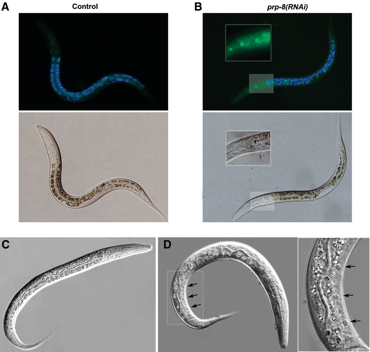
egl-1::GFP expression is ectopically induced in prp-8(RNAi) animals. Expression of the transgene opIs56 [Pegl-1::2xNLS::GFP] after 24 h at 20°C in (A) control RNAi (empty vector) and (B) prp-8(RNAi). Size of the animals and the germline development stage indicate that in our experimental conditions prp-8(RNAi) worms develop to a similar stage as N2 during the first 24 h. prp-8(RNAi) worms ectopically express GFP in additional cells, including hypodermal seam cells (magnified area). Blue fluorescence is shown to label areas with autofluorescence. Images displayed are representative of three different experimental replicates. (C,D) Animals treated with prp-8(RNAi) display additional apoptotic cell corpses. The ced-6 mutant larvae treated with (C) control RNAi and (D) prp-8(RNAi). Black arrows indicate apoptotic cells (button-like refractile corpses) that were found in prp-8(RNAi) worms but not in control animals. The right panel shows a magnified image of the area highlighted with a white box.




