


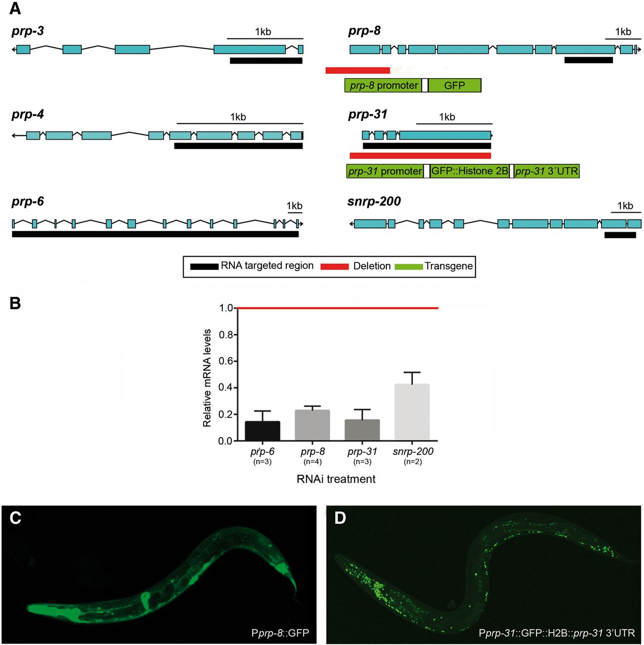
C. elegans toolkit to study s-adRP. (A) Scheme of s-adRP genes in C. elegans including the regions that are targeted by RNAi clones (black bars), the deleted fragment in the prp-31(gk1094) and prp-8(gk3511) alleles (red bars), and the elements of the prp-8 and prp-31 reporters generated in this study (green rectangles). The prp-31(gk1094) allele consists of a 5-bp insertion/1953-bp deletion. prp-8(gk3511) is a 1823-bp deletion. (B) Quantification of prp genes’ expression levels after their respective inactivation by RNAi. mRNA levels of prp genes are represented relative to the expression in gfp(RNAi) control animals (arbitrary value of 1, indicated with a red line). Transcript levels are normalized against tbb-2 levels in each case. RNA for analysis was obtained from up to four biological replicates (n). RNA from samples used for RNA-seq analyses were included. Error bars represent standard error of the mean. (C) Representative confocal image showing a transgenic worm expressing the transgene sEx12486, which consists of the GFP under the control of a promoter region for prp-8. (D) Representative confocal image showing a transgenic worm expressing the transgene cerEx79, which consists of the fusion protein GFP::H2B under the control of prp-31 promoter and 3′UTR.




