


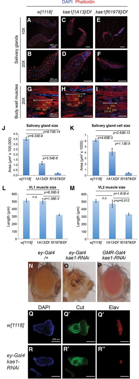
Differential requirements of kae1 in mitotic vs. nonproliferating tissues. Larval tissues were analyzed from kae1[1A13] and kae1[f01978] hemizygotes, which fully lack imaginal discs (Fig. 6). (A–I) Immunostaining of dissected tissues, using DAPI (blue) and phalloidin (red). (A–F) Salivary glands (and some attached fat bodies) imaged at 10× to visualize entire glands (A–C) and at 20× to emphasize individual cell sizes (D–F). (G–I) Body wall musculature. Selected muscles VL1-4 and LO1 are labeled. The length of a body segment is denoted with the double arrow lines. (J) Quantifications of salivary gland sizes (n = 10 all genotypes) and (K) salivary gland cell sizes (n = 15 cells from the distal tip measured in each of eight glands, all genotypes). Although kae1[1A13]/Df larvae exhibit severe defects in mitotic tissues, the overall salivary gland size and cell size are more modestly affected. The null allele kae1[f01978] is severely compromised in both aspects of the salivary gland. (L) Quantifications of VL1 muscle length and (M) VL2 muscle length. kae1[1A13]/Df larvae exhibit normal muscle sizes, while the null allele is only ∼40% reduced. Error bar shows standard error; all statistical analyses were unpaired Student's t-tests. (N–P) Adult eyes bearing Gal4 driver alone (N) or expressing kae1-RNAi under control of ey-Gal4 (O) or GMR-Gal4 (P). The former is active in the proliferating eye primordium, whereas the latter is active in post-mitotic photoreceptor neurons. (Q,R) Third instar eye-antennal imaginal discs stained for DAPI, Cut (green, to label cone cells) and Elav (red, to label photoreceptors). Compared to the control disc (Q), depletion of kae1 using ey-Gal4 induces severely defective eye development. All scale bars, 200 μm.




