


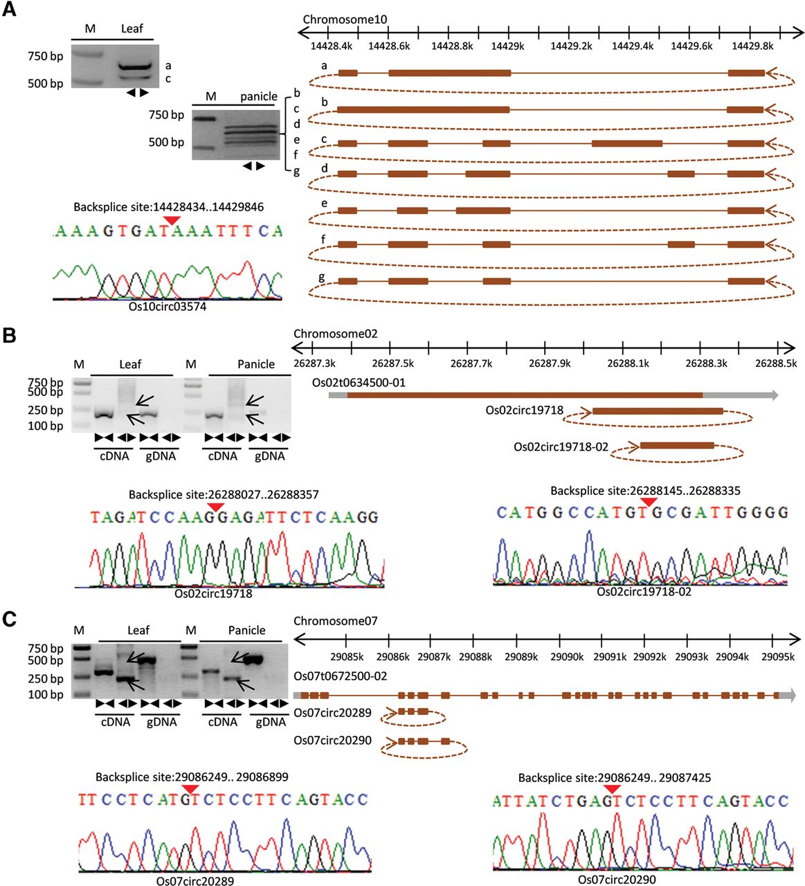
Visualization and validation of three rice alternative circularization events. (A) In the upper left, the PCR amplifications with divergent primers of circRNA “Os10circ03574” in leaf (two bands are indicated) and panicle (six bands are indicated) were performed. In the lower left, the backsplice site was further confirmed by Sanger sequencing. The sequence was visualized using a raw trace file. Red inverted triangles indicate the junction loci. On the right, seven alternative splicing circularization structures of “Os10circ03574” obtained from Sanger sequencing are shown in detail. A brown bar denotes an exon, a gray bar denotes UTR, and a brown line denotes an intron. (B,C) Similar to A, the results from PCR amplification and Sanger sequencing are displayed. Alternative backsplicing circularization events were identified and validated.




