


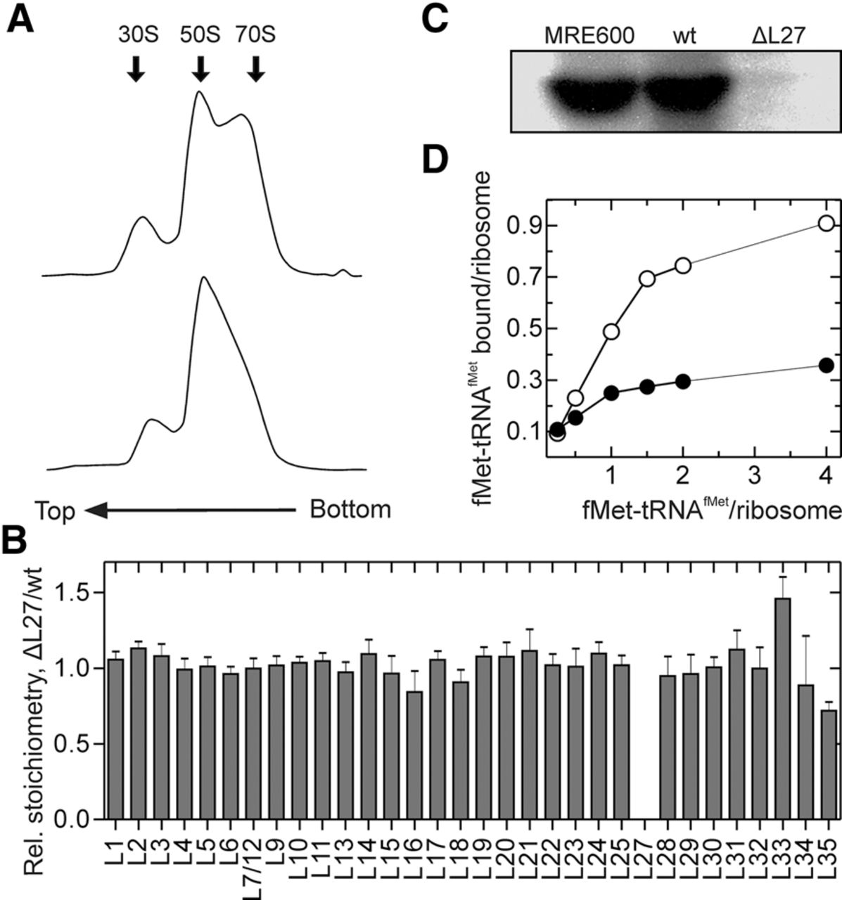
Characterization of ΔL27 ribosomes. (A) Sucrose-gradient centrifugation profile of wt (upper panel) and ΔL27 (bottom panel) ribosomes at 5 mM Mg2+. (B) Quantification of ribosomal proteins by mass spectrometry. The ratio of the average protein concentrations (as defined by label-free quantification) ΔL27/wt was plotted. Error bars represent the standard deviation of four technical replicates. (C) Western blot of ribosomal proteins from MRE600, wt, and ΔL27 ribosomes using anti-L27 antibody. (D) Determination of the active concentration of ΔL27 ribosomes. The extent of initiation was determined by the radioactivity retained on nitrocellulose filters after incubation of wt (open circles) or ΔL27 (closed circles) ribosomes with mRNA, initiation factors, GTP, and increasing concentrations of f[3H]Met-tRNAfMet.




