


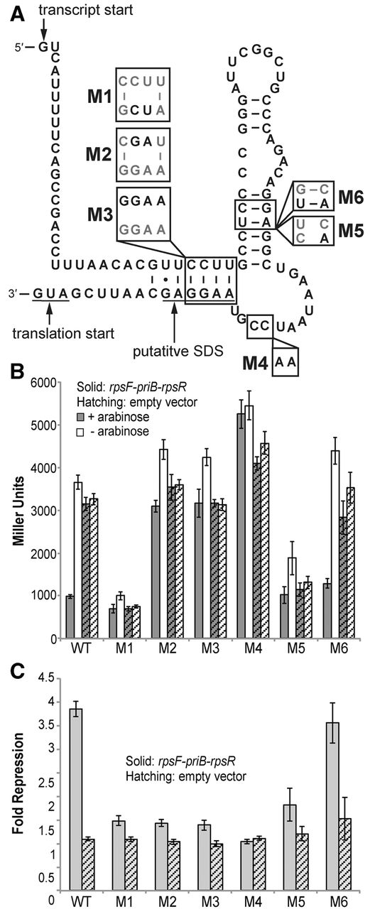
FIGURE 2.
(A) The presumed secondary structure of the rpsF 5′ UTR used for reporter studies with mutations M1–M6. The transcription start site (Maciag et al. 2011), translational start, and putative Shine–Dalgarno (SD) sequence are indicated. (B) β-galactosidase activity of cells carrying plasmids with the unmutated rpsF_leader (WT) or each mutant RNA (M1–M6) and the rpsF-priB-rpsR overexpression plasmid or the empty vector (pBAD33) under induced and uninduced (±arabinose) conditions. (C) Fold repression as calculated in Figure 1C of the unmutated rpsF_leader (WT) and each mutant RNA (M1–M6).




