


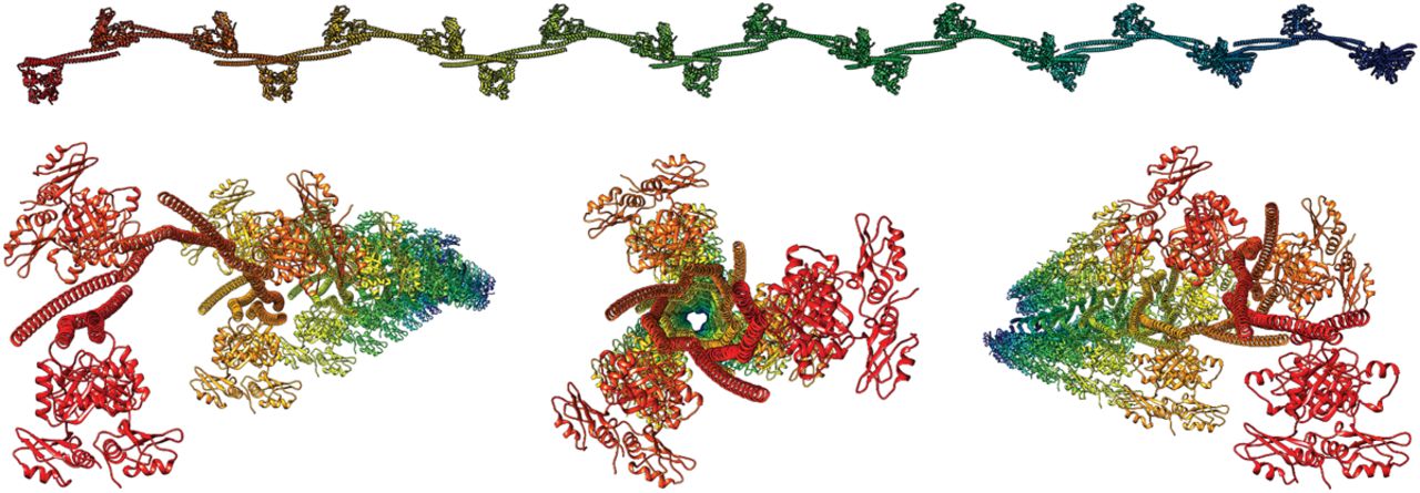
FIGURE 3.
Different views of a large paraspeckle protein multimer model containing 22 core dimers and colored according to chain ID. The modeled arrangement results in a spiraling relative orientation of the subsequent tandem RRM regions. Figures prepared with Chimera (Pettersen et al. 2004).




