


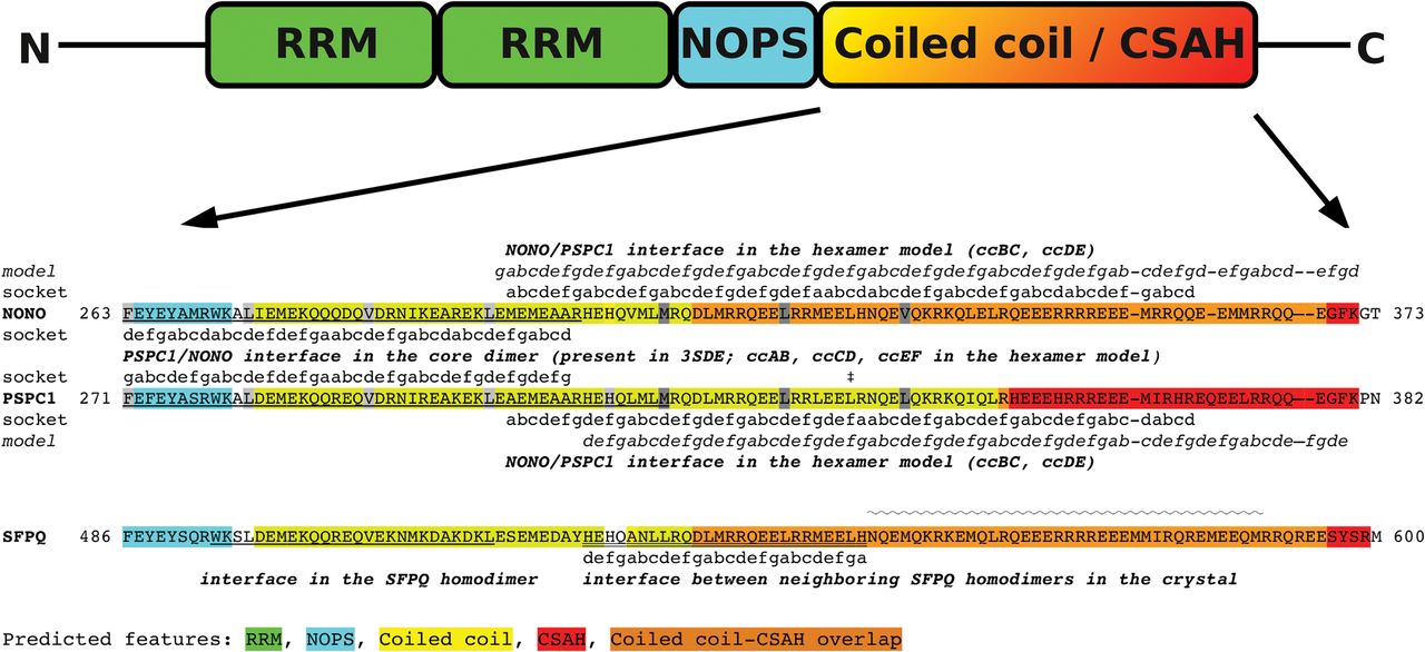
Partial sequence alignment of human core paraspeckle proteins with predicted, observed, and modeled features. Start and end positions of the segments shown are marked. The coiled-coil segment identified by SOCKET in the PSPC1/NONO dimer structure (3SDE) is underlined and the heptad positions are indicated (labels between the two sequences). Truncation before the residue marked with “‡” results in the loss of PSPC1 localization to paraspeckles (Passon et al. 2012). Hydrophobic residues in the hendecad repeat are highlighted in light and dark gray for the crystallized and the extended regions, respectively. In the case of PSPC1 and NONO, the heptad positions considered during model building and those obtained by SOCKET for the generated hexamer are shown (labels “model” and “socket”, above and below the NONO and PSPC1 sequences, respectively). For SFPQ, residues involved in coiled-coil formation of the dimer are underlined (based on analysis of the 4WIJ structure, note that the coiled coil is not symmetric despite being homodimeric, i.e., not all underlined residues participate in the coiled coil in both subunits). Residues identified to form the interdimer left-handed coiled coil are underlined with a double line and the heptad positions are indicated (Lee et al. 2015). The segment forming the extended single α-helix in the 4WIJ crystal structure is marked with a wave.




