


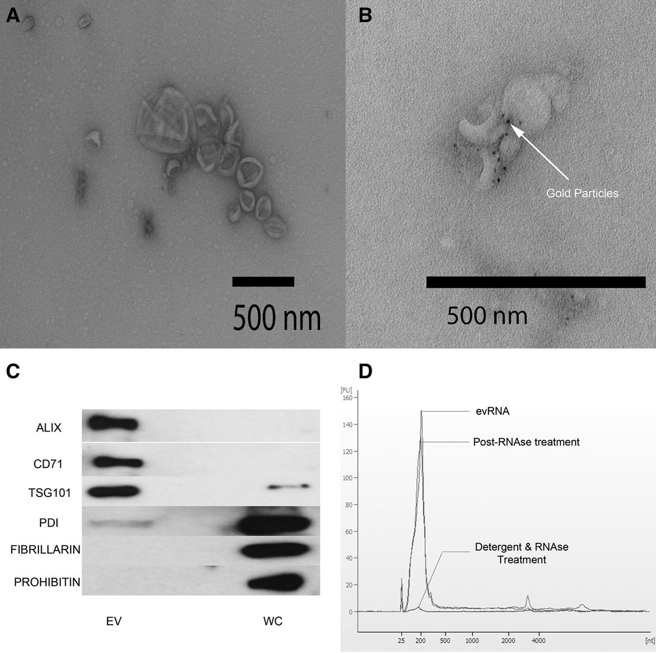
Validation of purification of extracellular vesicles (EVs). (A) Transmission electron microscopy image of K562 EVs after negative staining shows classic cup-shaped vesicles that are on average smaller than 200 nm. (B) Immuno-electron microscopy image of purified EVs labeled with anti-CD81 (mouse mAb) and detected by goat anti-mouse IgG secondary conjugated with 5 nm gold. Dark spots on the image are the electron dense gold elements conjugate to IgG secondary antibody. (C) Bioanalyzer RNA profile (RNA Pico-chip) of untreated EVs (red), RNA profile of EVs treated with RNase (green), and RNA profile of EVs treated with detergent and RNase (blue). (X-axis) Nucleotide lengths; (y-axis) fluorescent units. (D) Western blot analysis of proteins from K562 EVs and whole cell. Proteins selected for detection were previously identified to be enriched in EV or whole cell. (EV enriched) ALIX (PDCD6IP gene), CD71 (TfR1 gene), TSG101 (TSG101 gene). (Whole cell) PDI (PDI gene), FIBRILLARIN (FBL gene), PROHIBITIN (PHB gene).




