


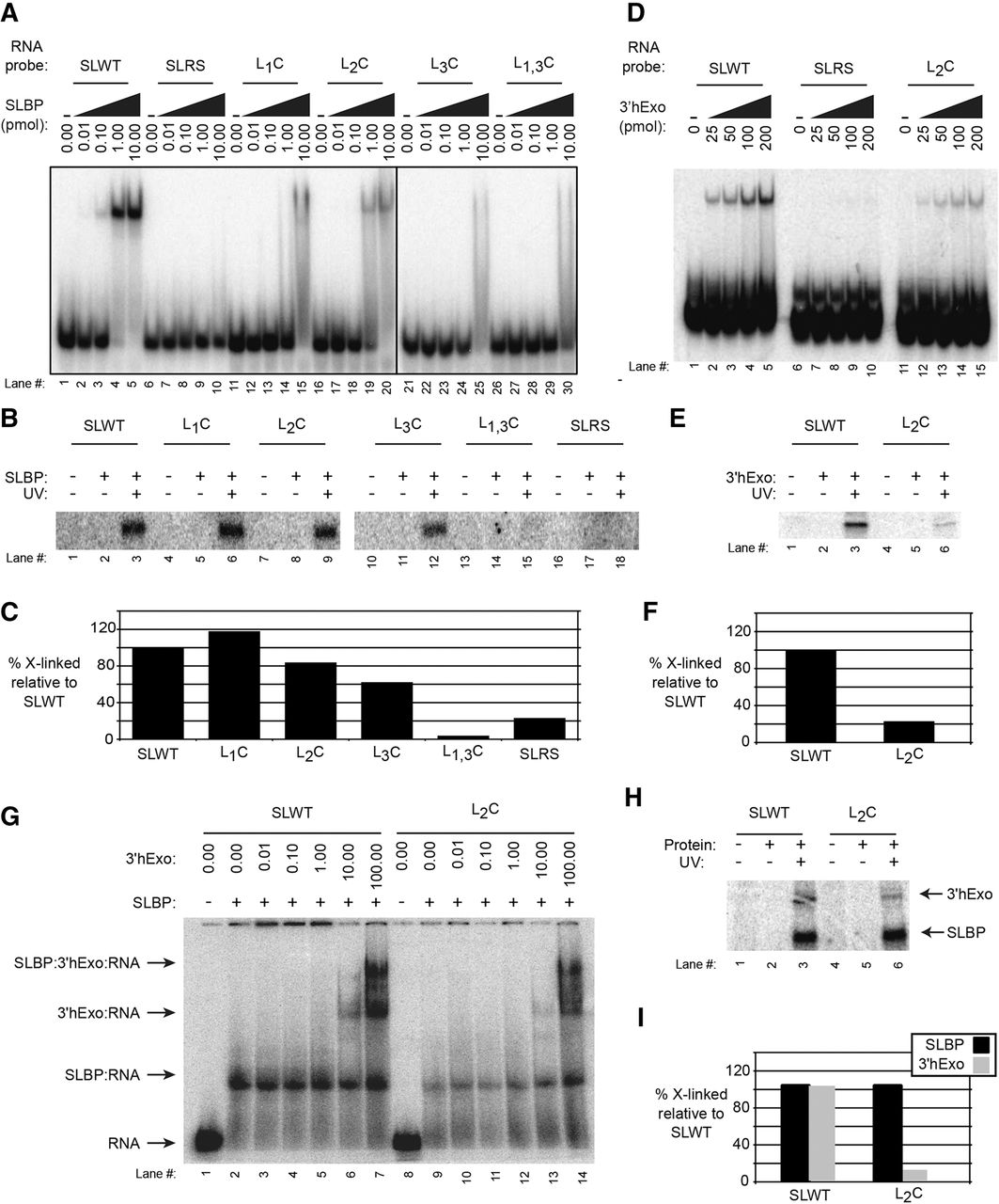
SLBP crosslinks to the L1,3U and the 3′hExo crosslinks primarily to L2U of the loop. (A) Electrophoretic mobility shift assays (EMSAs) were performed to ascertain binding of SLBP to mutant SL sequences. Cytosines were substituted for specific uridines in the loop and the effect on SLBP binding determined by EMSA. A stem–loop reverse sequence (SLRS) that does not bind SLBP was used as a negative control. The position(s) of the mutation(s) in the variant SL probes are indicated: L1C changes the first position in the loop from U to C, L2C changes the second, L3C changes the third, and L1,3C changes both the first and third positions from U to C. (B,C) The ability to crosslink recombinant SLBP to these probes was determined by UV irradiation followed by SDS-gel electrophoresis. The relative crosslinking is shown in the graph. (D) The binding of 3′hExo to the SL wild type (SLWT) (lanes 1–5), SLRS (lanes 6–10), and L2C (lanes 11–15) was measured by EMSA. (E) We carried out in vitro UV crosslinking with the SLWT (lanes 1–3) and L2C (lanes 4–6) probes. (F) The results were quantified by PhosphorImager. (G) We incubated the SLWT (lanes 1–7) and L2C probes (lanes 8–15) with 10 pmol of the SLBP RNA-processing domain and increasing amounts of 3′hExo. A ternary complex was formed in similar amounts with both probes. (H) The complexes with SLWT (lanes 1–3) and the L2C probes (lanes 4–6) were crosslinked with UV light, and the two proteins resolved by SDS-gel electrophoresis. (I) The intensity of crosslinking quantified on a PhosphorImager.




