


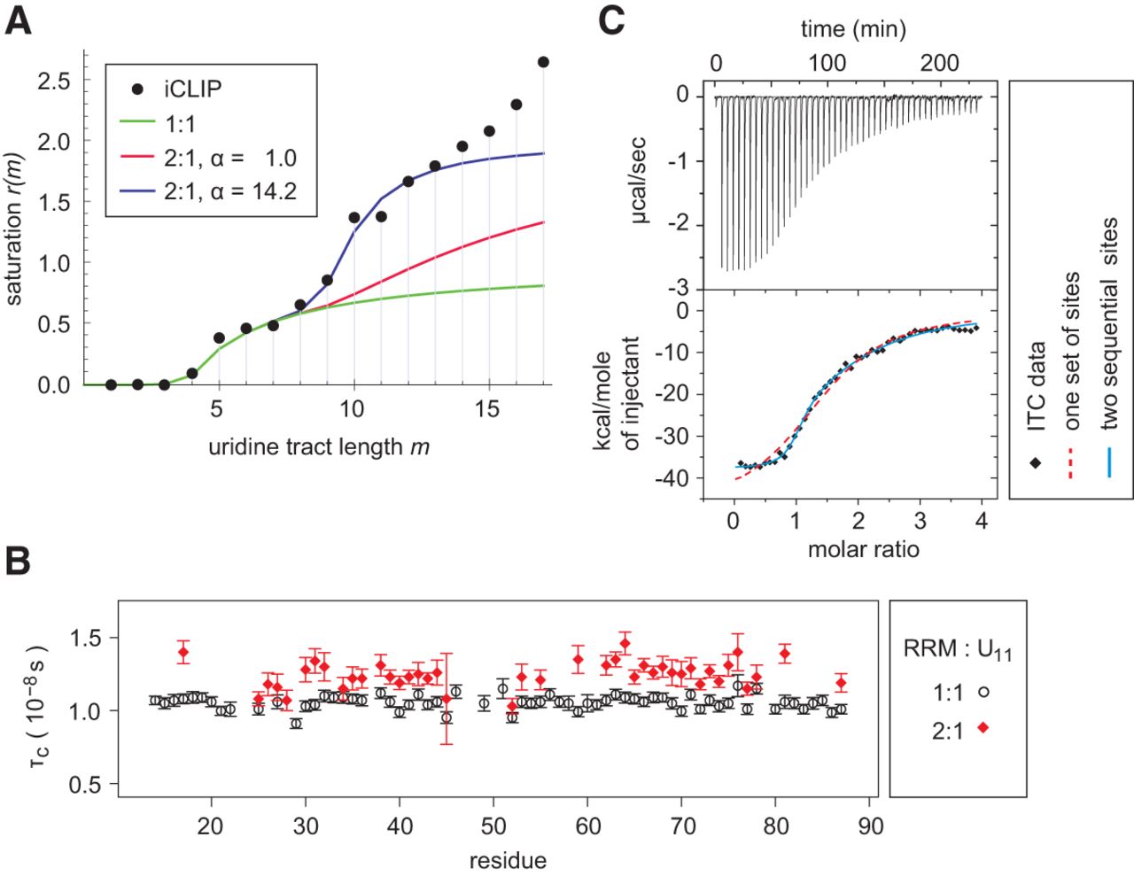
(A) Occupancy of cellular uridine tracts by hnRNP C. Experimental hnRNP C cross-link saturation (König et al. 2010) is compared with a model allowing only 1:1 RRM:RNA stoichiometry (in green), a model assuming two independent RRM ligands binding one uridine tract (in red), and a model allowing ligand cooperativity (in blue). In all three cases the scaling factor (see Materials and Methods) s = 73. (B) Rotational correlation times τC obtained from resolved amide resonances of residues 14–87 of hnRNP C RRM in presence of one or half equivalent of (U)11 RNA. (C) ITC of the RRM binding to (U)11 oligomer (320 μM of protein in the syringe, 17 μM of RNA in the cell, 6 μL/inj.). Experimental isotherm is fitted to two binding models.




