


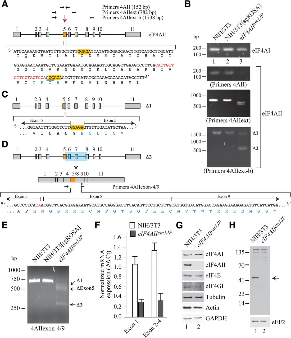
(A) Generation of NIH/3T3 eIF4AIIem1JP cells. Schematic diagram illustrating the exon/intron structure of the murine eIF4AII gene and denoting exon 5 (orange shading; downward arrow) targeted by Cas9 for deletion mutagenesis. The positions of the primers used to characterize the eIF4AII locus following CRISPR/Cas9-mediated gene editing are shown. The red sequence denotes the region to which the Cas9 sgRNA was designed and the underlined 5′-GGG-3′ triplet indicates the position of the PAM motif. The orange highlighted sequence denotes a duplicated region that likely led to microhomology-mediated NHEJ to generate Δ1 (see text for details). The TPGR motif 1b is shown in green. (B) PCR analysis of eIF4AIIem1JP. DNA was isolated from parental NIH/3T3 cells (lane 1), NIH/3T3 cells expressing Cas9 and an sgRNA targeting the neutral Rosa26 locus (Malina et al. 2013) (lane 2), or eIF4AIIem1JPcells (lane 3) and used in PCRs with the indicated primer pairs (Table 1). PCR products were analyzed on 1% agarose gels. Products were excised from the gel and analyzed by direct sequencing. (C) Schematic diagram representing configuration of the Δ1 eIF4AII allele in eIF4AIIem1JP. (D) Schematic diagram representing configuration of the Δ2 eIF4AII allele in eIF4AIIem1JP. (E) RT-PCR analysis of mRNA isolated from the indicated cell lines. Complementary DNA was generated as indicated in Materials and Methods using an eIF4AII-specific primer. PCRs were performed using the indicated primer pairs and products analyzed on a 1.2% agarose gel. Products were excised from the gel and analyzed by direct sequencing. (F) RT-qPCR analysis of eIF4AII mRNA in NIH/3T3 and eIF4AIIem1JP cells. Expression of eIF4AII was calculated relative to GAPDH levels and is based on ΔΔCT values. (G) Western blot probing of extracts prepared from NIH/3T3 (lane 1) or eIF4AIIem1JP (lane 2) using antibodies directed to the proteins shown to the right. (H) Western blot probing of extracts prepared from NIH/3T3 (lane 1) or eIF4AIIem1JP (lane 2) using an N-terminal directed antibody (ab31218, Abcam). Arrow indicates position of migration of eIF4AII.




