


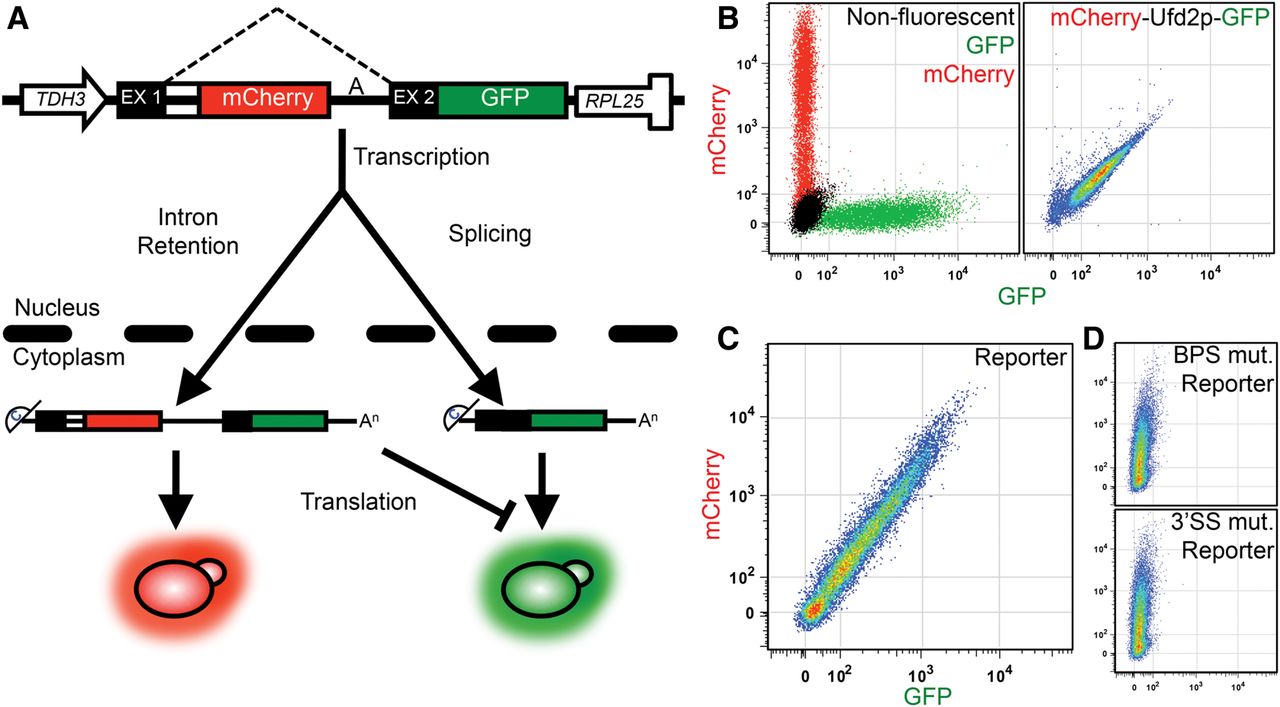
Gene expression reporter design and validation. Design (A) of the gene expression reporter construct is shown. Expression of the synthetic reporter was driven by the strong constitutive TDH3 promoter. Exon 1 (EX1), the intron, and exon 2 (EX2) were designed using features of the inefficiently spliced CYH2 gene. Dashed lines indicate the region comprising the intron. The mCherry ORF was placed in frame with EX1 and the eGFP ORF was cloned in frame with EX2. Exported intron-containing pre-mRNAs and mature mRNAs are translated resulting in proteins that fluoresce red and green, respectively. The construct was cloned into pRS316 (see Supplemental Material for more detail). Control samples (B) for flow cytometry are shown. Phenographs for wild-type yeast harboring an empty vector (non-fluorescent), or vectors expressing GFP-only or mCherry-only are shown (left). (Right) A phenograph of the expression of the mCherry-Ufd2p-GFP dimer results in a 1:1 mCherry:GFP signal. The pseudo color represents cell density within the phenograph. Reporter expression in wild-type yeast (C) is displayed and referred to as a phenograph. Phenographs for branch point (A to G) and 3′ splice site (CAG to CuG) mutant reporters (D) in wild-type yeast are shown (top and bottom, respectively). The y- and x-axis depict mCherry and GFP fluorescence, respectively.




