


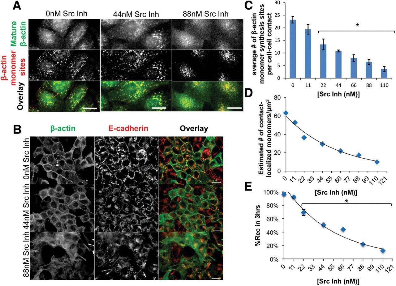
Inhibiting Src activity decreases contact-localized β-actin monomer synthesis to perturb epithelial adherens junction assembly. (A) De-convolved epifluorescence images of MDCK cells 3 h after the return to Ca2+ recovery media without (0 nM Src Inh) or with (44 nM Src Inh and 88 nM Src Inh) Src-1 inhibitor. β-actin monomer synthesis sites are in red and mature β-actin is in green. Scale bars, 10 μm. (B) De-convolved epifluorescence images of MDCK cells 3 h post-Ca2+ experiment illustrating β-actin (green) and E-cadherin (red) without (0 nM Src Inh) or with (44 nM Src Inh or 88 nM Src Inh) Src-1 inhibitor. Scale bars, 20 μm. (C) Graph of the average number of β-actin monomer synthesis sites per cell contact. (D) Dose-dependent curve shows the effects of increased concentrations of Src-1 inhibitor on the estimated number of contact-localized β-actin monomers. Scale bars, 10 μm. (E) Dose-dependent curve showing the relationship between increased Src-1 inhibitor on the %Rec of MDCK monolayer in 3 h after a Ca2+ experiment. Error bars mean ± SEM based on five cells in three independent experiments. (*) P < 0.05.




